Download
1 / 18
200 likes | 419 Vues
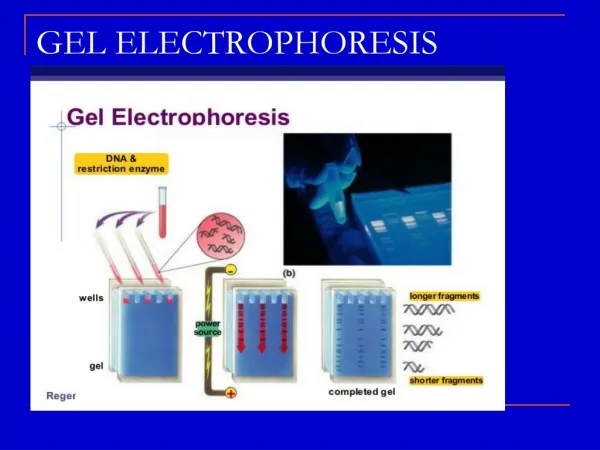
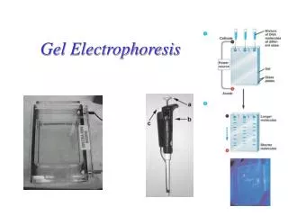
Gel Electrophoresis. Method for separating molecules (DNA, proteins, etc.) on the basis of physical or chemical properties such as: (1) size (2) shape (3) electrical charge. Gel electrophoresis. A method of separating DNA in a gelatin-like material using an electrical field

E N D
Gel Electrophoresis Method for separating molecules (DNA, proteins, etc.) on the basis of physical or chemical properties such as: (1) size (2) shape (3) electrical charge
Gel electrophoresis • A method of separating DNA in a gelatin-like material using an electrical field • DNA is negatively charged • when it’s in an electrical field it moves toward the positive side DNA – + “swimming through Jello”
Electrophoresis of DNA • Gels are made of agarose or polyacrylamide • DNA samples loaded, voltage applied • Negatively charged DNA migrates toward “+” electrode • Smaller DNA fragments migrate faster
A Typical Electrophoresis Gel Setup Negative end- - - - - - - - - - - - - Direction of DNA movement Direction of DNA movement Positive end+ + + + + + + + + +
Background About Agarose • Agarose is a mixture of long chains of saccharides (sugar) • It is extracted from a seaweed and is used in oriental cuisine as a gelling agent for desserts • The greater the agarose concentration, the smaller the pores within the gel • This changes the ease with which the DNA can travel through the gel
+ + + + + + + + + + + + + + + + + + + + + + + + ++ + + + + + + + + + DNA is negatively charged and therefore repelled from the negative pole and attracted towards the positive pole - - - - - - - - - - - - - - - - - - - - - - - - - - - - - - - - - - -
+ + + + + + + + + + + + + _ _ _ _ _ _ _ _ _ _ _ _
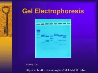
A Typical Image of an Agarose Gel Under UV Light Largest DNA fragments Decreasing DNA Size • The DNA fragments can be visualized using a special dye that specifically binds DNA and fluoresces under illumination with UV light Smallest DNA fragments
Sizing The Fragments of DNA • The sizes of the various fragments can be identified by including a “ladder” in the gel • A ladder is a mixture of DNA fragments of known size • When run in a gel electrophoresis, these fragments will separate into distinct bands that can be used as references
Typical Ladders-100 bp & 1 kbp (1000 bp) • The 100 bp ladder is composed of a mixture of small fragments (100 to 2000 bp) • The 1000 bp ladder is composed of a mixture of larger fragments (500 to 12000 bp)
Sizing a Gel Product Base Pairs (bp) 4000 3000 2000 1600 1000 500 2000 bp 1000 bp 1Kbp Sample ladder
What is the Size of this Fragment? 4000 3000 2000 1600 1000 500 Sample 1Kbp ladder
Procedure for Day 1 • Determine amount of agarose and buffer needed to make 50 ml of 1% agarose. • Microwave the mixture for about 30 seconds. • Using a hot pad, take the flask out of the microwave and swirl gently. Make sure all the powder has dissolved. • Set on your bench to cool for 5 minutes. • Insert the comb into the middle slot on the gel tray. • Pour the cool to the touch liquid gel into the tray. • Let the gel harden for at least 15-20 minutes.
Procedure for Day 2 • Pull out the comb from the wells by pulling gently upwards. • Place the gel into the gel box. Make sure the wells are at the black end. • Add 350 ml of TAE buffer to flood the gel and fill the wells. • Load 7 µl of each sample into a well on the gel. • During the lab, note where each sample was loaded on a diagram of your gel.
Put the cover on the electrophoresis box so that the electrodes are connected. • Plug the other end of the electrodes into the power supply and run at 100 volts for 30 minutes. • After the samples have run out, make a drawing of how the gel looked in your lab notebook.
Conclusion • What is the charge of these colored dyes? • What would happen to the samples if you ran the gel at 200 volts for 30 minutes? • What is the charge on DNA?
Which colored dye moved the farthest distance through the gel? • Which colored dye moved the shortest distance through the gel? • Which colored dye is the smallest in size? • Which colored dye is the largest in size? • What is the relationship between distance traveled and size of the molecule in an agarose gel?