
The Respiratory System
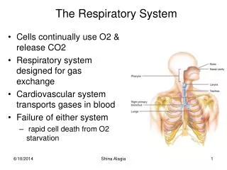
The Respiratory System. Cells continually use O2 & release CO2 Respiratory system designed for gas exchange Cardiovascular system transports gases in blood Failure of either system rapid cell death from O2 starvation. Respiratory System Anatomy. Nose Pharynx = throat Larynx = voicebox

The Respiratory System
E N D
Presentation Transcript
The Respiratory System • Cells continually use O2 & release CO2 • Respiratory system designed for gas exchange • Cardiovascular system transports gases in blood • Failure of either system • rapid cell death from O2 starvation Shina Alagia
Respiratory System Anatomy • Nose • Pharynx = throat • Larynx = voicebox • Trachea = windpipe • Bronchi = airways • Lungs - upper respiratory tract is above vocal cords • lower respiratory tract is below vocal cords Shina Alagia
External Nasal Structures • Skin, nasal bones, & cartilage lined with mucous membrane • Openings called external nares or nostrils Shina Alagia
Nose -- Internal Structures • Large chamber within the skull • Roof is made up of ethmoid and floor is hard palate • Internal nares are openings to pharynx • Nasal septum is composed of bone & cartilage • Bony swelling or conchae on lateral walls Shina Alagia
Functions of the Nasal Structures • Olfactory epithelium for sense of smell • Pseudostratified ciliated columnar with goblet cells lines nasal cavity • warms air due to high vascularity • mucous moistens air & traps dust • cilia move mucous towards pharynx • Paranasal sinuses open into nasal cavity • found in ethmoid, sphenoid, frontal & maxillary • lighten skull & resonate voice Shina Alagia
Pharynx • Muscular tube (5 inch long) hanging from skull • skeletal muscle & mucous membrane • Extends from internal nares to cricoid cartilage • Functions • passageway for food and air • resonating chamber for speech production • tonsil (lymphatic tissue) in the walls protects entryway into body • Distinct regions -- nasopharynx, oropharynx and laryngopharynx Shina Alagia
Nasopharynx From internal nares to soft palate openings of auditory (Eustachian) tubes from middle ear cavity adenoids or pharyngeal tonsil in roof Shina Alagia
Oropharynx From soft palate to hyoid bone fauces is opening from mouth into oropharynx palatine tonsils found in side walls, lingual tonsil in tongue Common passageway for food & air Shina Alagia
Laryngopharynx Extends from hyoid bone to cricoid cartilage Common passageway for food & air & ends as esophagus inferiorly Shina Alagia
Cartilages of the Larynx • Thyroid cartilage forms Adam’s apple • Epiglottis---leaf-shaped piece of elastic cartilage • during swallowing, larynx moves upward • epiglottis bends to cover glottis • Cricoid cartilage---ring of cartilage attached to top of trachea • Pair of arytenoid cartilages sit upon cricoid • many muscles responsible for their movement • partially buried in vocal folds (true vocal cords) Shina Alagia
Larynx • Cartilage & connective tissue tube • Anterior to C4 to C6 • Constructed of 3 single & 3 paired cartilages Shina Alagia
Vocal Cords • False vocal cords (ventricular folds) found above vocal folds (true vocal cords) • True vocal cords attach to arytenoid cartilages Shina Alagia
Trachea • Extends from larynx to T5 anterior to the esophagus and then splits into bronchi • Layers • mucosa = pseudostratified columnar with cilia & goblet • submucosa = loose connective tissue & seromucous glands • hyaline cartilage = 16 to 20 incomplete rings • open side facing esophagus contains trachealis m. (smooth) • internal ridge on last ring called carina Shina Alagia
Trachea and Bronchial Tree • Full extent of airways is visible starting at the larynx and trachea Shina Alagia
Histology of the Trachea • Ciliated pseudostratified columnar epithelium • Hyaline cartilage as C-shaped structure closed by trachealis muscle Shina Alagia
Bronchi and Bronchioles • Primary bronchi supply each lung • Secondary bronchi supply each lobe of the lungs (3 right + 2 left) • Tertiary bronchi supply each bronchopulmonary segment • Repeated branchings called bronchioles form a bronchial tree Shina Alagia
Histology of Bronchial Tree • Epithelium changes from pseudostratified ciliated columnar to nonciliated simple cuboidal as pass deeper into lungs • Incomplete rings of cartilage replaced by rings of smooth muscle & then connective tissue • sympathetic NS & adrenal gland release epinephrine that relaxes smooth muscle & dilates airways • asthma attack or allergic reactions constrict distal bronchiole smooth muscle Shina Alagia
Pleural Membranes & Pleural Cavity • Visceral pleura covers lungs --- parietal pleura lines ribcage & covers upper surface of diaphragm • Pleural cavity is potential space between ribs & lungs Shina Alagia
Gross Anatomy of Lungs • Base, apex (cupula), costal surface, cardiac notch • Oblique & horizontal fissure in right lung results in 3 lobes • Oblique fissure only in left lung produces 2 lobes Shina Alagia
Mediastinal Surface of Lungs • Blood vessels & airways enter lungs at hilus • Forms root of lungs • Covered with pleura Shina Alagia
Structures within a segment of Lung • Branchings of single arteriole, venule & bronchiole are wrapped by elastic CT • Respiratory bronchiole • simple squamous • Alveolar ducts surrounded by alveolar sacs & alveoli • sac is 2 or more alveoli sharing a common opening Shina Alagia
Cells Types of the Alveoli • Type I alveolar cells • simple squamous cells where gas exchange occurs • Type II alveolar cells (septal cells) • free surface has microvilli • secrete alveolar fluid containing surfactant • Alveolar dust cells • wandering macrophages remove debris Shina Alagia
Alveolar-Capillary Membrane • Respiratory membrane = 1/2 micron thick • Exchange of gas from alveoli to blood • 4 Layers of membrane to cross • alveolar epithelial wall of type I cells • alveolar epithelial basement membrane • capillary basement membrane • endothelial cells of capillary Shina Alagia
Details of Respiratory Membrane • Find the 4 layers that comprise the respiratory membrane Shina Alagia
Double Blood Supply to the Lungs • Deoxygenated blood arrives through pulmonary trunk from the right ventricle • Bronchial arteries branch off of the aorta to supply oxygenated blood to lung tissue • Venous drainage returns all blood to heart • Pulmonary blood vessels constrict in response to low O2 levels so as not to pick up CO2 on there way through the lungs Shina Alagia
Respiration • Respiration is exchange of primarily oxygen and carbon dioxide between atmosphere and human body Shina Alagia
Respiration: Steps • Respiration is achieved in four steps • Pulmonary ventilation: Inspiration + Expiration • External respiration: Diffusion across alveolar-capillary membrane • Gas transport: Transport of O2 and CO2 • Internal respiration: Exchange between ICF and tissue capillary Shina Alagia
Breathing or Pulmonary Ventilation • Air moves into lungs when pressure inside lungs is less than atmospheric pressure • How is this accomplished? • Air moves out of the lungs when pressure inside lungs is greater than atmospheric pressure • How is this accomplished? • Atmospheric pressure = 1 atm or 760mm Hg Shina Alagia
Boyle’s Law • As the size of closed container decreases, pressure inside is increased • The molecules have less wall area to strike so the pressure on each inch of area increases. Shina Alagia
Dimensions of the Chest Cavity • Breathing in requires muscular activity & chest size changes • Contraction of the diaphragm flattens the dome and increases the vertical dimension of the chest Shina Alagia
Quiet Inspiration • Diaphragm moves 1 cm & ribs lifted by external intercostal muscles • Intrathoracic pressure falls and 2-3 liters inhaled Shina Alagia
Quiet Expiration • Passive process with no muscle action • Elastic recoil & surface tension in alveoli pulls inward • Alveolar pressure increases & air is pushed out Shina Alagia
Intra-pleuralPressures Helps keep parietal & visceral pleura stick together and alveoli inflated • Always subatmospheric (756 mm Hg) • As diaphragm contracts intrapleural pressure decreases even more (754 mm Hg) Shina Alagia
Summary of Breathing • Alveolar pressure decreases & air rushes in • Alveolar pressure increases & air rushes out Shina Alagia
Pressure changes during Pulmonary Ventilation • Intrapulmonary pressure =atmospheric(760) • Intrapleural pressure = sub-atmospheric (-4) • During inspiration: Intrapleural Pressure= -6 mm of Hg Intrapulmonary pressure = -1 mm of Hg • During expiration: Intrapleural pressure= -3 mm of Hg Intrapulmonary pressure = +1 mm of Hg Shina Alagia
Compliance of the Lungs • Ease with which lungs & chest wall expand depends upon 1. Elastic recoil of lungs & 2. surface tension • Some diseases reduce compliance • tuberculosis forms scar tissue • pulmonary edema --- fluid in lungs & reduced surfactant Shina Alagia
Alveolar Surface Tension • Thin layer of fluid in alveoli causes inwardly directed force = surface tension • water molecules strongly attracted to each other • Causes alveoli to remain as small as possible • Detergent-like substance called surfactant produced by Type II alveolar cells • lowers alveolar surface tension Shina Alagia
Pneumothorax • Pleural cavities are sealed cavities not open to the outside • Injuries to the chest wall that let air enter the intrapleural space • causes a pneumothorax • collapsed lung on same side as injury • Atelectasis: Collapsing of lung due to lack of sufficient surfactant Shina Alagia
Airway Resistance • Resistance to airflow depends upon airway size(diameter) • increase size of chest • airways increase in diameter • contract smooth muscles in airways • decreases in diameter Shina Alagia
Lung Volumes and Capacities • Tidal volume = amount air moved during quiet breathing • MVR= minute ventilation is amount of air moved in a minute • Reserve volumes ---- amount you can breathe either in or out above that amount of tidal volume • Residual volume = 1200 mL permanently trapped air in system • Vital capacity & total lung capacity are sums of the other volumes Shina Alagia
External AND Internal Respiration Shina Alagia
Dalton’s Law • Each gas in a mixture of gases exerts its own pressure • as if all other gases were not present • partial pressures denoted as p • Total pressure is sum of all partial pressures • atmospheric pressure (760 mm Hg) = pO2 + pCO2 + pN2 + pH2O • to determine partial pressure of O2-- multiply 760 by % of air that is O2 (21%) = 160 mm Hg Shina Alagia
Henry’s Law • Quantity of a gas that will dissolve in a liquid depends upon the amount of gas present and its solubility • Breathing O2 under pressure dissolves more O2 in blood • Clinical application of Henry’s law: Hyperbaric Oxygenation • Use of pressure to dissolve more O2 in the blood • treatment for patients with anaerobic bacterial infections (tetanus and gangrene) • Used to treat heart disorders, carbon monoxide poisoning, cerebral edema, bone infections, crush injuries Shina Alagia
Mechanism of external respiration • Mechanism: Diffusion • Driving force: Partial pressure difference between alveoli and alveolar capillary • pO2 in alveoli>pO2 in alveolar capillary • pCO2 in alveoli<pCO2 in alveolar capillary • Constant adding of O2 and removal of CO2 keeps it that way. Shina Alagia
External Respiration • Exchange of gas between air & blood • Gases diffuse from areas of high partial pressure to areas of low partial pressure • Deoxygenated blood becomes oxygeneted • Compare gas movements in pulmonary capillaries to tissue capillaries Shina Alagia
Rate of Diffusion of Gases • Factors that influence diffusion are: • Partial pressure of gases in air • p O2 at sea level is 160 mm Hg • 10,000 feet is 110 mm Hg / 50,000 feet is 18 mm Hg • Large surface area of our alveoli • Diffusion distance is very small • Solubility & molecular weight of gases • O2 smaller molecule diffuses somewhat fast • CO2 dissolves 24X more easily in water so net outward diffusion of CO2 is much faster Shina Alagia
Internal respiration • Driving force: Diffusion due to partial pressure difference • Mechanism: Similar to external respiration only directions of gas movements are changed • pO2 in systemic capillary>pO2 in ISF • pCO2 in systemic capillary <pCO2 in ISF Shina Alagia
Internal Respiration • Exchange of gases between blood & tissues • Conversion of oxygenated blood into deoxygenated • Observe diffusion of O2 inward • at rest 25% of available O2 enters cells • during exercise more O2 is absorbed • Observe diffusion of CO2 outward Shina Alagia
Gas Transport Shina Alagia
Oxygen Transport in the Blood • Oxyhemoglobin contains 98.5% chemically combined oxygen and hemoglobin • inside red blood cells • Does not dissolve easily in water • only 1.5% transported dissolved in blood • Only the dissolved O2 can diffuse into tissues • Factors affecting dissociation of O2 from hemoglobin are important Shina Alagia


