Download
1 / 10
120 likes | 468 Vues
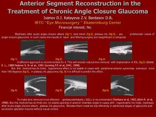
Normal Anterior Chamber Angle. Schwalbe’s line. Schlemm’s canal. Narrow Anterior Chamber Angle. Angle Scans. Scleral spur (red arrow) Schlemm’s canal (blue arrow) Schwalbe’s line (green arrow). Closed angle with peripheral anterior synechiae. OD. OS. Normal cornea.

E N D
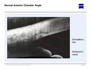
Normal Anterior Chamber Angle Schwalbe’s line Schlemm’s canal
Angle Scans • Scleral spur (red arrow) • Schlemm’s canal (blue arrow) • Schwalbe’s line (green arrow)
Normal cornea Structures visualized: Epithelium (red arrow), Bowman’s membrane (green arrow), Stroma (blue arrow), Descemet’s and endothelium (yellow arrow)
LASIK One month post-op LASIK flap edge visible in scan
Corneal Scar • 50 y/o male, bungee cord injury 2 months prior • Traumatic corneal ulceration • Thinned cornea with epithelial defect. • Tear film visible over defect.
Descemet’s Stripping Endothelial Keratoplasty (DSEK) • Note gap between edge of DSEK and Descemet’s membrane of recipient