
Problem 1
E N D
Presentation Transcript
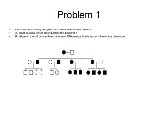
Problem 1 • Mutation in noncoding sequence change the number of protein molecules produce, but, generally each protein molecule made will have a normal amino acid sequence. Give an example of a mutation that agrees with the rule. Give examples of some exceptions to this rule, and describe how the alteration in the amino acid sequence are generated. Problem 8 Chapter 11 text
Answer Problem 1 • Give an example of a mutation that agrees with the rule. • many simple beta-thalassemia with transciption mutations • fragile X syndrome • Give examples of some exceptions to this rule, and describe how the alteration in the amino acid sequence are generated. • exceptions to this rule can arise, for example, from splice site mutations that lead to missplicing of an exon. The exon may be excluded from the mRNA, generating either an in-frame deletion of the protein sequence or causing a change in the reading frame, leading to the inclusion of different amino acids in the protein sequence
bs globin gene GTG 5’ 3’ Region recognized by probe normal bA globin gene GTG 5’ 3’ Region recognized by probe Problem 2 • Sickle cells anemia was one of the first diseases for which a unique molecular marker was discovered. Sickle-cell Anemia is due to a single-nucleotide substitution (A T) in the second position of the sixth codon of the beta-globin gene that is responsible for the difference between the standard A and sickle-cell S alleles. The sequence of the standard A allele (CCTGAGG) happends to correspond to an MstII restriction site (CCTNAGG) that is not present in the S allele (CCTGTGG). The beta-globin gene region includes two flanking MstII sites (red lines). • a. For allele A how many bands will total DNA from the individual tested produce when cut with MstII • b. For allele S how many bands will total DNA from the individual tested produce with cut with MstII • c. Indicate the geneotype for each individual • d. Indicate individual who have the sickle cell trait • e. Which child has the highest fitness rating in a malaria ridden area in Africa
Answer Problem 2 • a. For allele A how many bands will total DNA from the individual tested produce when cut with MstII • two bands • b. For allele S how many bands will total DNA from the individual tested produce with cut with MstII • one band • Total DNA from the individual tested is cut with MstII: a probe specific for the region including the nucleotide substitution will then produce two bands if the standard A allele is present, and one band if the sickle-cell S allele is present; a heterozygote (sickle-cell trait) will therefore show three bands. • c. Indicate the geneotype for each individual • allele A two small bands • allele S one large band • d. Indicate individual who have the sickle cell trait • people who have the trait are heterozygous. Heterozygous individuals will have three bands • Individuals I-1,1-2,II-3 • e. Which child has the highest fitness rating in a malaria ridden area in Africa • individual II-3 heterozygous survive malaria better • Note this test depends on the coincidence that the nucleotide substitution responsible for sickle-cell happens to occur in such a way as to create an RFLP: the MstII site itself has nothing to do with sickle-cell anemia.
Problem 3 The figure below shows a PCR bases micro-array hybridization assay of the DMD exons. In the figure below PCR-fragments containing DMD exons are spotted in triplicate on each array (top left exons 1-24, top right exons 25-48, bottom left exons 49-72, bottom right exons 73-79). Top: signal obtained with control DNA (Cy-5 labeled). Bottom: signal obtained with DNA from a patient (Cy-3 labeled). Does this patient have DMD. If they do what exons are deleted
Answer Problem 3 Does this patient have DMD. Yes . If they do what exons are deleted. The assay suggests they are missing exon 3-20 missing 3-20 control