Tooth Transplantation
Tooth Transplantation. Tooth Transplantation. a viable alternative endodontic treatment or extraction ? fixed or removable prosthetic treatment is not ideal treatment in adolescent. Why tooth transplantation is successful?. Hertwig’s epithelial root sheath. Tooth transplantation.
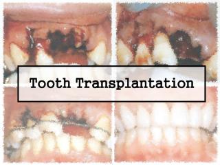

Tooth Transplantation
E N D
Presentation Transcript
Tooth Transplantation • a viable alternative • endodontic treatment or extraction ? • fixed or removable prosthetic treatment is not ideal treatment in adolescent
Why tooth transplantation is successful? Hertwig’s epithelial root sheath
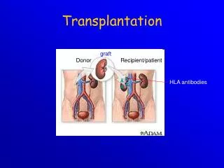
Tooth transplantation • Autogenous transplantation • Allogeneic transplantation • Isogeneic transplantation • Xenegeneic transplantation
Indication • Sufficientcrown space and alveolar bone • No periapical or periodontal inflammation • Proximity of the transplant to the socket wall to assure rapid organization of the clot between the alveolar bone and the tooth • Excellent oral hygiene, low caries index
Tooth autotransplantation • Transplantation from one region to another • Transalveolar transplantation Sagne S. : Autotransplantation of teeth Int Dent J. 1985 : 35 , 280-283
Transplantation from one legion to another • Transplant from lower 3rd molar to lower 1st molar • Transplant from upper 3rd molar to upper 1st molar • Transplant from lower premolar to upper premolar or upper premolar to lower premolar • Transplant from premolar to upper central incisor
Transplant from lower3rd molar to lower 1st molar • Most reported cases of autotransplantation • a result of caries the first molar is frequently missing or removal in adolescents • Developing third molars are usually available in adolescents • In a majority of case the rate of success is well over 95%* for an extended period of time *Andreasen et al. 1992
Indication for 3rd molar to 1st molar • Appropriate candidates are adolescent patients ( 13 to 20 years ) • Recent lost or about to lose a permanent first molar • A suitable third molar • The roots on the donor tooth developed to the point of bifurcation ( should be equal to approximately 3 -4 mm in root length ) • A fully formed crown
Indication for 3rd molar to 1st molar • A suitable third molar • Complete enamel calcification coincides closely with development of the bifurcation • The third molar should be no larger than the first molar it is replacing • Slight of third molar to make it a suitable size is acceptable • During instrumentation in the removal of the donor tooth, the vulnerable tooth buds are avoided
Surgical technique 1.Mobilization of the transplant • A mucoperiosteal flap prepared by a sulcular incision from the mesial of second premolar to the distal of second molar then extending distolaterallyand no vertical incision, assuring an excellent blood supply • It is important that the flap design allow both adequate surgical field and blood supply
Surgical technique 1.Mobilization of the transplant • The impacted third molar is carefully exposed , avoiding any contact of bone-cutting instruments with the tooth, grasping the crown with forcepsavoid trauma to the root sac
Surgical technique 1.Mobilization of the transplant • The tooth is then luxated, elevated from its position and gently returned to its position or maintained in its socket • Leaving the donor tooth in the socket after luxation will allowit to continue to receive nutrients and be hydrate while the host site is being preparation
Surgical technique • Preparation of the host site • The first molarand interradicular bone are carefully removed • Amount of cortical bone removed is critical, if an injudicious amount of bone is removed , there will not be an adequate bone support
Surgical technique • Preparation of the host site • The required amount of bone can be estimated by radiograph • Irrigate and inspected for debris before a trial positioning of the transplant
Surgical technique • Transplantation and stabilization • The third molar is carried forward to new socket • The area of resistance is relieved before seating the third molar • The transplant may be stripped to seating but the roots ofthe transplantation should not be scraped or filed
Surgical technique • Transplantation and stabilization • The occlusion should be carefully examined to be certain that the opposing teeth will not exert pressure on the transplant • avoid premature contact • Infraocclusion about 2 – 3 mm
Surgical technique • Transplantation and stabilization • Mucoperiosteal flap is repositioned and suture • The transplant is splinted in position using 0.14 gauge stainless steel wire • The wire ligation technique used can be figure eight or circumferential technique
Surgical technique • Transplantation and stabilization • Additional stabilization of the transplant can be achieved by gently packing periodontal surgical dressing such as Coe-Pak around the transplant and adjacent teeth
Postoperative care and follow-up • Postoperative instructions are the same as those given following extraction of impacted teeth • After surgery 1 day : the tooth has retained its new position : periodontal pack still in good position : swelling is within acceptable levels 7 days : stitch off
Postoperative care and follow-up • The patient should be seen at weekly intervals • At the end of a month the transplant may still be slightly mobile, but splinting can be removed • Follow-up every month within 6 months • every 3 monthwithin 2 years • every year
Postoperative care and follow-up • At each visit should be checked • The stability of the transplant • Sulcular depth • Gingival recession • Vitality test • Occlusion • Root formation, thickness of periodontal ligament, root resorption in radiograph • Oral hygiene
Precautions • The precaution that help ensure successful autogenous transplantation are the following : • Root development of donor tooth is between 1/3 to 1/2 of the total root • Hertwig’s epithelial root sheath is not injured during surgery • The host site is prepared to avoid injury to the epithelial root sheath • The patient should be healthy with adequate oral hygiene • Pulpy foods that might pack into the cervicular space should be avoided • The patient should consider the procedure important, keep operative site clean and avoid trauma from occlusion
Transplantation from one legion to another • Transplant from lower 3rd molar to lower 1st molar • Transplant from upper 3rd molar to upper 1st molar • Transplant from lower premolar to upper premolar or upper premolar to lower premolar • Transplant from premolar to upper central incisor
Surgical technique The treatment plan is to transplant a maxillary second premolarto the maxillary central incisor which is to be removed due to root resorption
Surgical technique The maxillary central incisor is extracted
Surgical technique The socket is enlarged with surgical bur The socket is expanded palatally , then rinse with saline
Surgical technique Testing the size of socket by a glass replica of a premolar
Surgical technique Removing maxillary second premolar using gentle luxation movement
Surgical technique Repositioning of the transplant, it is placed 45◦ rotate in order to achieve sufficient cervical width
Surgical technique Splint the transplant with 0.20 mm stainless steel wire
Surgical technique Complete treatment, after grinding and restore with crown
Summary Although it is not possible to perform tooth autotransplantationin all patients with nonrestorable molars, it may be a viable alternative in some instance
Reference Bowden David E. J. et al : Autotransplantation of premolar teeth to replace missing maxillary central incisor, British Journal of orthodontics, Vol. 17, 1990 Munksgaard : Text book and color atlas of traumatic injuries to the teeth, 1994 Plainfield S. et al : A viable alternative : Tooth transplantation, Journal of Prosthodontics, Vol. 50, 1983 Robison J. Peter and Grossman I. Louis : Tooth Transplantation, Clinical transplantation in dental specialties Smith J. J. et al : Successful Autotransplantation, Journal of Endodontics, Vol.13, 2, 1987