Download
1 / 34
370 likes | 741 Vues
Infections of the lower respiratory tract. Elizabeth Wasserman Clinical Microbiologist, Pathcare Laboratories Extraordinary professor, Division of Medical Microbiology, Stellenbosch University. Lower respiratory tract: distal to the larynx .

E N D
Infections of the lower respiratory tract Elizabeth Wasserman Clinical Microbiologist, Pathcare Laboratories Extraordinary professor, Division of Medical Microbiology, Stellenbosch University
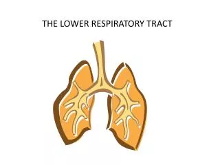
Lower respiratory tract: distal to the larynx This environment is sterile under normal conditions. NATURAL HOST DEFENSES Mucus Cilia Phagocytosis by polymorphs and macrophages Lysozyme Interferon Secretory IgA Bronchus associated lymphoid tissue
CONDITIONS THAT MAY DISRUPT NATURAL HOST DEFENSES Viral infections Underlying lung conditions e.g. chronic bronchitis Smoking Iatrogenic interventions e.g. intubation and ventilation
Microbiology of relevant pathogens S. pyogenes S. pneumonia covered in previous lectures H. influenza M. catarrhalis
Infection with the so-called ‘atypicals’: • Mycoplasmapneumoniae • Chlamydophylapneumoniae • Legionella
Mycoplasma pneumonia: • Part of the historically labelled atypical pneumonias • Epidemiology: infection occurs world-wide. Preponderance of infection in late summer and early autumn. Spread is fostered by close contact, e.g. in a family. Children are infected more often than adults. Pneumonia is less frequent as a consequence of infection with increasing age, but severity increases with the age of the patient.
Clinical features: • insidious onset with malaise, myalgia, sore throat, headache. • Dry cough starts later. • Patients usually do not appear seriously ill and few warrant admission. • Radiographic evidence of pneumonia often appears before physical signs. • The course of the disease is variable, but may extend over several weeks. • Relapse is frequent.
A prolonged paroxysmal cough simulating the features of whooping cough may occur in children. • Very severe infections may occur in adults, usually in those with immunodeficiency or sickle cell anaemia. • Extra pulmonary manifestations: Stevens-Johnson syndrome and other rashes; arthralgia; meningitis or encephalitis; haemolytic anaemia (brought about by cold agglutinins); myocarditis and pericarditis
Laboratory diagnosis: • Cold agglutinins
Molecular methods is the diagnostic approach of choice, when available. However, asymptomatic carriage of the organism occur and PCR may be positive in the absence of active disease • Serological tests: IgG peaks around 3 – 4 weeks following infection and persists for years. Therefore, a high titre is required for diagnosis of active infection, or a 4x rise in titre . IgM more reliable in children, IgA useful all ages.
Direct antigen detection • Isolation of the organism by culture is impractical as it takes long (up to 3 weeks or longer) and is not available in all laboratories.
Treatment: • Intrinsically resistant to beta-lactams (absence of peptidoglycan target) • Treatment of choice: Tetracyclines, erythromycin, new fluoroquinolones. • Therapy should be started based on clinical suspicion, and a 3 week course is justified, particularly with laboratory confirmation
Chlamydophilapneumoniae • Can infect humans and animals • Can only replicate intracellularly • Airborn transmission occurs as reticulate bodies.
C. pneumoniae causes pneumonia, pharyngitis, bronchitis, otitis and sinusitis with an incubation period of about 21 days. • Studies have shown it to be the third or fourth most common cause of pneumonia following S. pneumonia and H. influenza. • Recurrences are frequent, affecting different areas in the lung.
Serological link suggested between C. pneumoniae and coronary heart disease, atherosclerosis and sarcoidosis. The organism can also be demonstrated in atheromatous plaques.
Legionella • L. pneumophilais a facultative intracellular bacterium that can invade and replicate inside amoebae in the environment, which can thus serve as a reservoir for L. pneumophila, as well as provide protection from environmental stresses, such as chlorination.
Epidemiology: • Only acute bacterial pneumonia which may occur in outbreak form. • Bacteria disseminated in aerosols which may travel for 1-2km from the source. • Ponds in cooling towers of refrigeration plants in air-conditioning systems • Domestic hot water systems in hotels and hospitals • Warm water in nebulisers and oxygen line humidifiers • Whirlpool spa baths and showers
Infection with Legionella pneumophilia Legionellae give rise to two main clinical syndromes: • Legionnaires’ disease: may progress rapidly and extend to involve two or more lobes of the lung. Incubation period is 2-10 days. Characterized by high fever and symptoms such as respiratory distress, cough, confusion and focal neurological signs. Significant mortality in untreated patients with underlying lung disease • Pontiac fever: brief febrile influenza-like illness with no mortality.
Legionellae survive in free-living amoebae, and may be protected from drying and disinfectants in the cysts of such amoebae. • Control: hot water should be heated above 60°C before distribution, and should not lie stagnant and cooling in the pipes. • Cold water should be kept below 20°C and not allowed to stagnate. • Constant chlorination may suppress growth.
Laboratory diagnosis: • Gram stains not useful, except to demonstrate the presence of other pathogens. • Blood cultures not useful • Immunofluorescent staining with specific monoclonal or polyclonal antisera can be done on respiratory secretions, pleural fluid or lung biopsy. • Cultures require special media • Antigen tests in urine is rapid and specific • Serology: AB take at least 8 days to develop. Paired sera NB to show development of AB or rise in titre. Some patients may not develop AB for some weeks or, rarely, for several months. AB may persist for months or years.
Infective Disease Complexes of the Lower Respiratory Tract • Acute tracheo – bronchitis • Pneumonia • Lung abscess • Empyema • Infection in the presence of underlying disease: Exacerbation of chronic bronchitis Bronchiectasis Cystic fibrosis
Acute tracheo – bronchitis Etiology: • Most commonly caused by viruses (rhinovirus, influenza or para-influenza) • Secondary bacterial infections may follow: • H. influenzae • S. pneumoniae • S. aureus (rare)
Pneumonia Etiology: • Community vs. hospital acquired • Fungal pneumonia • Parasitic lung infections
Clinical clues to the possible cause of a pneumonia History: • Community or hospital acquired? • Age of your patient • Underlying disease • Possible aspiration • Local epidemiology • Travel • Occupation • Exposure to animals • Air-conditioning cooling towers
Community or hospital acquired? Community acquired pneumonia: Route of infection: endogenous infection • S. pneumoniae • S. aureus • H. influenzae • Klebsiella pneumoniae • Tuberculosis • Viruses
Hospital acquired pneumonia: Route of infection may be either endogenous or exogenous. The same list as community acquired pneumoniae, but now also: • Gram negative bacteria including Pseudomonas auruginosa, Acinetobacter sp. and others that may be more resistant to antimicrobials
Lung abscess • S. aureus • Klebsiella sp. • Anaerobes • Entamoeba histolytica
Exacerbation of chronic bronchitis Ethiology: • Viral (rhino en para-influenza viruse) • Secondarybakterial infection: • H. influenzae • S. pneumoniae • K. pneumoniae • Pseudomonas • M. catarrhalis
Empyema • S. pneumoniae • S. aureus • Anaerobes • Gram negative bacilli • S. millerigroup • Mycobacteriae
Bronchiectasis Secondary infection due to: • Haemophilusinfluenzae • Streptococcus pneumoniae • Anaerobes e.g. Bacteroides • Pseudomonas aeruginosa • Klebsiella pneumoniae • Aspergillosis
Cystic fibrosis Secondary infection due to: • In kids: S. aureus • In adults: Pseudomonas aeruginosa Resistance to antimicrobials and the growth of organisms in biofilms makes treatment difficult.