Environment
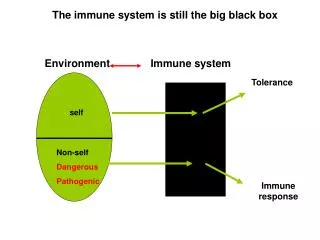
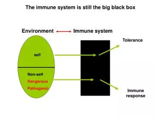
The immune system is still the big black box. Environment. Immune system. Tolerance. self. Non-self Dangerous Pathogenic. Immune response. V i rus. 3 hrs. Diversity Fast development. 3 hr s. PATHOGENS HAVE SHORT GENERATION TIME. PATOGENS. Bact eria. Vi ruses. parasites.

Environment
E N D
Presentation Transcript
The immune system is still the big black box Environment Immune system Tolerance self Non-self Dangerous Pathogenic Immune response
Virus 3 hrs Diversity Fast development 3 hrs PATHOGENS HAVE SHORT GENERATION TIME PATOGENS Bacteria Viruses parasites
Development, education Humans have longer generation time, Need a sophysticated protection system Birth Reproducrion at 35
INHERITED (PRIMARY) Loss of function mutation of genes of the immune system Enhanced susceptibility to infections Particular types of pathogens depending on the gene defect Did not show up until 1950 - antibiotics ACQUIRED Due to infectious diseases AIDS Other virus infections Malnutrition Artificial immunosuppression Drugs Radioactive irradiation IMMUNODEFICIENCIES AGE AND HEALTH DEPENDENT IMMUNOSUPPRESSIVE DRUGS
PRIMARY IMMUNODEFICIENCIES • MOST ARE RECESSIVE MUTATION OF SINGLE GENES • Dominant traits have been eliminated from the population • Autosomal genes • Disease in homozygous children • Heterozygous children are carriers • X-linked genes • Single gene defect causes disease in males • Single gene defect in females renders the affected woman carrier • Mutation in the IFNγ receptor results in binding without intracellular signaling - dominant DISSEMINATED INFECTION BY THE BCG STRAIN OF Mycobacterium USED FOR VACCINATION
The impact of recessive and dominant mutations in the IFN-γ receptor on monocyte activation.
Numerous Immunodeficiency loci reside on the X chromosome CGD: Chronic Granulomatous Disease WAS: Wiscott-Aldrich Syndrome SCID: Severe Combined Immunodeficiency XLA: X-linked Agammaglobulinemia XLP: X-linked Lymphoproliferative Disease XLHM: X-linked Hyper-IgM Syndrome
ANTIBODY DEFICIENCY - recurrent sinopulmonary and GI infections beginning after 3-4 mo. B cell development (XLA, IgA deficiency) B – T cell collaborations CD40 ligand, hyper IgM T CELL DEFICIENCY - SCID, opportunistic infections beginning early in infancy T cell development IL-7/Jak3 RAG-1 RAG-2 Artemis Thymus epithelial cells DiGeorge syndrome Purin catabolism DNS repair enzyme defect MHC class II synthesis blockade TYPES OF INHERITED IMMUNE DEFICIENCIES
TYPES OF INHERITED IMMUNE DEFICIENCIES 2. • PHAGOCYTIC SYSTEM • CD18 (CR3, CR4, LFA1) • NADPH oxidase (CGD) • Vesicular fusion • COMPLEMENT SYSTEM • some infections, primarily with encapsulated organisms and Neisseriae • Soluble and membrane factors • C3 • C1 – C4 • Complement inhibitors
Immunodeficiencies caused by B-cell defects Approx. 70% of all IDs Late Manifestation (7-9 month) Increased sensitivity to: Encapsulated bacteria Streptococcus pneumoniae Haemophylus influenzae Infection by Enterovirus, parasites
Encapsulated bacteria resist ingestion by phagocytes unless they are recognized by antibodies that fix complement.
SERUM IG LEVELS AS THE DEVELOPMENT OF THE IMMUNE SYSTEM PROGRESSES
ANTIBODY DEFICIENCY INABILITY TO CLEAR EXTRACELLULAR BACTERIA • X-LINKED AGAMMAGLOBULINEMIA XLA (Bruton’s agammaglobulinemia) 1:200,000 Symptoms • First few months of life is relatively normal (maternal Ig) • Tonsils are small, lymph nodes are barely palpable • Recurrent infection of sinuses and of the middle ear. Pneumonia. • Pyogenic bacteria – permanent tissue demage caused by enzyme release from bacteria and phagocytes bronchiectasis, chronic lung disease • Haemophilusinfluenzae, Streptococcus pneumoniae, Staphylococcus aureus, • Oral polio vaccine disseminate and cause poliomyelitis • T-cell responses to intracellular bacteria is normal (mycobacteria) • Lack of mature B cells plasma cells in the periphery
ANTIBODY DEFICIENCY INABILITY TO CLEAR EXTRACELLULAR BACTERIA • X-LINKED AGAMMAGLOBULINEMIA XLA (Bruton’s agammaglobulinemia) Genetic defect • Mutation in the Bruton’s tyrosine kinase, essential for B cell activation and development • NO B CELLS IN THE PERIPHERY – block at pre-B cell stage • Carrier mother XXHEALTHY non-random inactivation of X in B cells • Son XY DISEASE Son XY HEALTHY • Increased susceptibility to bacteria and enteroviruses Treatment – monthly injections of Gamma glob. (IVIG)
In patients with X-linked agammaglobulinemia (XLA), B cells do not develop beyond the pre-B cell stage.
When a T-cell defect results in antibody deficiency X-LINKED HYPER IgM SYNDROME
DIMINISHED ANTIBODY PRODUCTION AS A RESULT OF INHERITED DEFECT OF T CELL HELP • HYPER IgM SYNDROME (XLHIM) Symptoms • Susceptibility to pyogenic bacteria/opportunistic infection • Sensitivity to pyogenic bacteria Haemophilusinfluenzae, Streptococcus pneumoniae, Staphylococcus aureus • opportunistic infections • No specific antibody response to T-dependent antigens • low IgG, IgA, IgE • No germinal center formation • No leukocytosis but neutropenia • No macrophage activation by T cells CD40 – CD40L • opportunistic infections • sores and blisters in the mouth and throat • injection of GM-CSF (GMCSF is produced by macrophages)
Lack of germinal centers in lymph nodes of X-linked Hyper-IgM syndrome patients
DIMINISHED ANTIBODY PRODUCTION AS A RESULT OF INHERITED DEFECT OF T CELL HELP • HYPER IgM SYNDROME XLHIM Genetic defect Defect of the DC40L membrane receptor gene • X-linked, disease in males Treatment – antibiotics, – monthly injections of Gamma glob. (IVIG) – injection of GM-CSF (neutropenia)
HYPER IgM SYNDROME (Autosomal) -Intrinsic B cell defect, activation induced deaiminase (AID) deficiency. Cytidine uridine conversion. -The enyme is involved in affinity maturation and Ig. class switch - Lack of opportunistic infections
SELECTIVE IgA DEFICIENCY • 1/800 • - Chronic lung disease, • - Tendency to develop respiratory and gastrointestinal allergies and autoimmunity • - Over 40% of patients have anti-IgA antibodies – blood products containing IgA can cause severe allergic response. -Some are related to MHC class III region
SEVERE COMBINED IMMUNODEFICIENCY SCID DEFECT IN T CELL FUNCTIONS T cells are involved in all aspects of adaptive immunity • Persistent and recurrent infections with a broader range of pathogens than patients with B cell deficiences • Neither T cell-dependent antibody response nor cellular immunity are functional • T-, B+ NK- SCID • T- B- NK+
X-LINKED SEVERE COMBINED IMMUNODEFICIENCY SCID (Over 50% of cases) Symptoms • Small body weight, failure to thrive • Persistent and recurrent infections with a broader range of pathogens than patients with B cell deficiences • Opportunistic infections (Candida albicans, Pneumocystis carnii pneumonia)
Candida albicans infection in children with SCID Normal SCID The Hart shadow is clearly visible In the absence of the thymus
SEVER COMBINED IMMUNODEFICIENCIES The SCID phenotype can be caused by various gene defects • X-SCID – The common γ-chain of interleukin receptors is mutated IL-7 receptor • Part of IL2,4,7,9, 15, 21 Receptor • Autosomal SCID – mutation of Jak3 kinase IL-7 receptor-mediated signaling • Defect in the catabolism of purin bases – autosomal (T- B- NK+) • Adenosine deaminase (ADA) mutation – mental retardation • Purin nucleotide phosphorilase (PNP) • Accumulation of purin metabolites • Highly toxiC for developing lymphocytes, • Mutation of RAG enzymes – autosomal (Omen syndrome T- B- SCID) • No or little somatic gene rearrangement (RAPIDLY FATAL) • No circulating peripheral lymphocytes or very narrow repertoire • Mutation of a DNA repair enzyme – autosomal • DNA-dependent protein kinase (DNA-PK) involved in the cleavage of hairpins in somatic gene rearrangement • Bare lymphocyte syndrome – inhibited MHC synthesis • No CD4+ T cell response • CIITA co-activátor, RFX promoter binding protein or other transcription factor mutation • DiGeorge szyndrome • Development of thymic epithelial cells is inhibited – T cell development is inhibited
SÚLYOS KOMBINÁLT IMMUNODEFICIENCIÁK A SCID fenotípust eltérő gén hibák okozhatják • X-SCID – Az interleukin receptorok közös γ-láncának hibája közös gamma lánc 55% totál, része az IL2,4,7,9, 15, 21 receptoroknak T- B+, NK-, a periférián a limfocitákgykorlatilag csak B sejtek • Jak3 kinázmutációja IL-7 receptor jelátvitel
SEVER COMBINED IMMUNODEFICIENCIES The SCID phenotype can be caused by various gene defects • Defect in the catabolism of purin bases – autosomal (T- B- NK+) • Adenosine deaminase (ADA) mutation – mental retardation • Purin nucleotide phosphorilase (PNP) • Accumulation of purin metabolites • Highly toxic for developing T lymphocytes, less toxic for developing B lymphocytes ADA conc. A tímuszban kb 10-szeres
Treatment: • Bone marrow transplantation, preferably from a histocompatible sibling • Gene therapy
Diagnosis? • Most often SCID patients are in critical condition when they come to the clinic. • Their life can not always be saved. Need to be „healed” before bone marrow • Transplantation. • Is it possible to identify SCIDs before they get sick? 21, 2010, the Advisory Committee on Heritable Disorders in Newborns and Children voted unanimously to add screening for Severe Combined Immune Deficiency or SCID – commonly known as bubble boy disease – to the core panel for universal screening of all newborns in the United States. On May 21, 2010 Kathleen Sebelius, Secretary of Health and Services announced the addition of Severe Combined Immunodeficiency (SCID) to the core panel of 29 genetic disorders as part of her recommendation to adopt the national Recommended Uniform Screening Panel. SCID is the first nominated condition to be added to the core panel of disorders. States and US Territories screening all newborns for SCID are: WI, MA, NY, CA, CT, MI, CO, MS, DE, FL, TX, MN, IA, PA, UT and OH. T-cell receptor excision circle assay has revolutionized early identification of infants with SCID or severe T-cell lymphopenia. TREC fragments are identified by QPCR
V4 V5 V3 V6 V2 V7 V8 V9 D J V1 Somatic recombination produces T-cell receptor excision circle TREC A TREC can be detected by QPCR Early detection with practically no false positive results Loop of intervening DNA is excised
Immunodeficiency caused by defects in B and T cell development
Wiskott-Aldrich syndrome WAS – X-linked • A disease of defective reorganization of the actin cytoskeleton • Symptoms • Thrombocytopenia, small platelet size (decreased production of platelets in B. Marrow, increased destruction in spleen) • Eczema • No antibodies to carbohydrate antigens (role for T cells?) • pyogenic and opportunistic infections • severe infection with varichella (chicken pox) and herpes simplex (impaired CD8+ T-cell response) • Rearrangement of cytoskeleton upon T cell activation in the polarized contact with B cells, macrophages and target cells • Low IgM high IgA, IgE serum levels • Pyogenic bacterial, and opportunistic infections • B cell lymphomas • Genetic defect • Mutation in the WAS protein (WASP) expressed in white blood cells and megakaryocytes • Treatment • Bone marrow transplantation
Wiskott-Aldrich syndrome WAS – X-linked Thrombocytopenia 40000 /μL + smaller thrombocytes Loss of microvilli on T cells in Wiskott–Aldrich syndrome. Scanning electron micrographs of normal lymphocytes (panel a) and lymphocytes from a patient with Wiskott–Aldrich syndrome (panel b). Note that the normal lymphocyte surface is covered with abundant microvilli, which are sparse or absent from the patient's lymphocytes. Photographs courtesy of Dianne Kenney.
Wiskott-Aldrich syndrome WAS Defective T/B communication Expressed in white blood cells and megakaryocytes
Capping of TCR is defective in WASP negative T cells resting anti CD3 treated T cells from wt-mice T cells from WASP-/- mice
DEFECTS IN PHAGOCYTE FUNCTION ENHANCED SUSCEPTIBILITY TO BACTERIAL INFECTIONS • DEFICIENCY OF CD18/LEUKOCYTE ADHESION (LAD) • Common β-subunit of CR3, CR4 and LFA-1 • Blocked phagocyte migration from blood to infection site • Inhibited uptake and degradation of opsonized bacteria • Persistant infection with extracellular bacteria • Pyogenic infections • Defect in wound healing, severe inflammation of the gums • Lethal within the first decade of life without bone marrow transplant Omphalitis in LAD I patient
DEFECTS IN PHAGOCYTE FUNCTION ENHANCED SUSCEPTIBILITY TO BACTERIAL INFECTIONS • CHRONIC GRANULOMATOUS DISEASE – CGD (1 million in the US) • Mutation of NADPH oxidase – any of the 4 subunits (gp91 X-linked) • No superoxid O2- radical antibacterial activity is compromised • Chronic intracellularbacterial or fungal infections – granuloma formation • Aspergilus pneumonia • IFN-gamma improves resistance. Mechanism?? • Defect of glucose-6-phosphate dehydrogenase and myeloperoxidase less severe phenotype • Diagnosis: NBT + PMA treatment of neutrophils. Lack • of blue colour in CGD CGD patient with skin infections due to Serratia marcescens
CHRONIC GRANULOMATOUS DISEASE – CGD Phox complex NBT staining of neutrophils Healthy Carrier CGD
MUTATION OR FUNCTIONAL INACTIVATION OF SOLUBLE COMPLEMENT PROTEINS RESULTS IN IMMUNODEFICIENCY Classical Lectin Alternative MBL C1Inh C1 B-factor MASP C4 D-faktor C2 Properdin HANE* C3 Neisseria-infection Pyogenic infections I-factor immune complex immune complex H-faktor disease disease C5 Pyogenic infections immune complex C6 disease C7 C8 Neisseria-infection C9 severe pyogenic infections *HANE - hereditary angioneurotic edema Stabilizes alternative C3 convertase
MUTATION OF MEMBRANE BOUND COMPLEMENT PROTEINS RESULTS IN IMMUNODEFICIENCY MIRL = CD59 DAF accelerates the decay of classical and alternative C3 convertase
DEFECTS IN COMPLEMENT COMPONENTS IMPAIR ANTIBODY RESPONSES ACCUMULATION OF IMMUNE COMPLEXES • DEFICIENCY OF C3 OR ITS ACTIVATION • Susceptibility to pyogenic bacteria – inefficient opsonization • DEFICIENCY OF C5-C9 • Neisseria – NO complement mediated lysis • DEFICIENCY OF EARLY C1-C4 • No C3b and C4b fragments No CR1-mediated erythrocyte transport of immune complexes • Accumulation of immune complexes in blood, lymph, extracellular fluid deposition in tissues tissue demage macrophage activation inflammation • DEFICIENCY IN COMPLEMENT INHIBITORY FACTORS • I factor – uncontrolled C3 C3b C3 depletion inefficient opsonization • Decay Accelerating Factor DAF or CD59 MAC inhibitor – autoimmune-like condition lysis of autologous erythrocytes paroxysmal nocturnal hemoglobulinuria • C1 inhibitor – uncontrolled activation of the classical pathway vasoactive C2 accumulation of fluid in tissues – epiglottal swelling may lead to death by suffocation