CRANIUM-SKULL
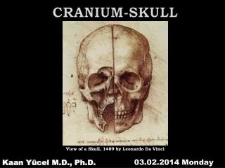
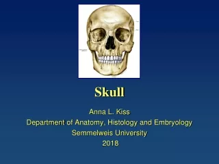
CRANIUM-SKULL. View of a Skull, 1489 by Leonardo Da Vinci. 03.02.2014 Monday. Kaan Yücel M.D., Ph.D. . SKULL. s keleton of the head. cranium 22 bones excluding ossicles of the ear. SUTURAE – SUTURES(S). . SKULL. M andible L ower jaw bone. . SKULL. s keleton of the head.


CRANIUM-SKULL
E N D
Presentation Transcript
CRANIUM-SKULL View of a Skull, 1489by Leonardo Da Vinci 03.02.2014 Monday Kaan Yücel M.D., Ph.D.
.SKULL skeleton of the head cranium 22 bones excluding ossiclesof the ear
.SKULL Mandible Lower jaw bone
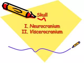
.SKULL skeleton of the head Neurocranium Viscerocraniumfacial skeleton
22 bones in thecranium 8 @ neurocranium Frontal bone- single Occipital bone- single Parietalbones- paired Temporalbones- paired Sphenoidbone- single Ethmoid bone- single
neurocranium 1roof (calvaria) 1 floorbase (base of the skull)basicranium
CALVARIA TEMPORAL BONES PARIETAL BONES parts of the singlefrontal, sphenoid, &occipital bones CRANIAL BASE mainly parts of the Sphenoid bone Temporalbones Occipital bone
Frontal Bone OS FRONTALE
Frontal Bone 1. Squamavertical portion region of the forehead 2. Orbitalportion frontal orbit/orbitafrontalishorizontal portion formation of roofs of the orbital & nasal cavities Squama
parIETAL BoneS OS PARIETALE Twoparietal bones unite and form the sides & roof of the cranium. Each bone is irregularly quadrilateral in form.
parIETAL BoneS OS PARIETALE Parietaleminence (tuber parietale) an elevationnearthecentre of theconvexandsmoothexternalsurface Superiorand inferior temporal lines twocurvedlinescrossingthemiddle of the bone in an archeddirection
temporalBoneS OS temporale Situatedat the sides and base of the skull. Contributesmost of the lower portion of lateral wall of the cranium.
temporalBoneS • 3parts • Squamous part • Tympanic part • Petromastoid part
Squamouspart of thetemporal bone large flat plate, forms the anterior &superior parts of the temporal bone contributes to lateral wall of the cranium articulates anteriorly with greater wing of the sphenoid bone superiorlywithparietal bone
Squamouspart of thetemporal bone Zygomaticprocess anterior bony projection from the lower surface of the squamous part of the temporal bone Zygomaticarch zygomaticprocess of thetemporalbone+temporalprocess of thezygomatic bone
Temporomandindibularjoint mandibular fossa concavedepressionlocatedinferiorly @ squamousportion Head of themandibleenters here! articular tubercle downward projection of the anterior border of the mandibular fossa
Tympanicpart of thetemporal bone justbelow the origin of the zygomatic process Externalacoustic opening (pore) entrance to the external acoustic meatus (canal) leads to the tympanic membrane (eardrum).
Mastoidpart of thetemporal bone most posterior part of the temporal bone continuous with squamous part anteriorly articulates with parietal bone superiorly occipital bone posteriorly.
Mastoidpart of thetemporal bone mastoid process on thelateralaspect, cone-shapedprojectionfromtheinferiorsurface mastoid notch medialaspect of themastoidprocess
Petrouspartof thetemporal bone lateral to the basilar part of the occipital bone between greater wing of the sphenoid anteriorly basilar part of the occipital bone posteriorly.
Petrouspart of thetemporal bone foramen lacerum apex of thepetrouspartforms one of the boundaries ofthisforamen. opening for the carotid canal largecircularopeningposterolateraltotheforamenlacerum
Petrouspart of thetemporal bone Jugularforamen large opening between the occipital bone & petrous vein draining the brain 3 of the 12 cranialnervespassthrough here (CNs 9-10-11)
Styloidprocess needle-shaped bone marking projects from the lower border of the temporal bone. anteromedial to the mastoid process point of attachment for numerous muscles and ligaments stylomastoidforamen Transmitsthenerveforthemuscles of theface Posteriortothebase of thestyloidprocess Between styloidprocess & mastoidprocess CN VII FACIAL NERVE
sphenoIDAL BONE OS sphenoIdale at the base of the skull in front of the temporal bones 6 basilar part of occipital bone median portion body two great and two small wings extending outward from the sides of the body two pterygoid processes project from it below.
sellaturcicae(L. Turkish saddle) saddle-like bony formation on the upper surface of the body of the sphenoid Anterior& posterior clinoid processes
sellaturcicae(L. Turkish saddle) Clinoidmeans «bedpost» 4processes (2anterior 2 posterior) surround hypophysial fossa “bed” of the pituitary gland like the posts of a four-poster bed.
sellaturcicaE(L. Turkish saddle) composed of three parts tuberculumsellae(horn of saddle) hypophysial fossa (pituitary fossa) dorsum sellae(back of saddle)
FORAMINA on each side of the body,4 foramina perforate the greater wings of the sphenoid • Superior orbital fissure • between the greater and the lesser wings • Foramen rotundum • posterior to medial end of the superior orbital fissure • Foramen ovale • posterolateralto the foramen rotundum • Foramen spinosum • posterolateralto the foramen ovale • ROLS
Pterygoid processes lateral medial pterygoidplates Pterygoid fossa
OCCIPITAL BONE • OS OCCIPITALE • at the back and lower part of the cranium foramen magnum cranial cavity communicates with the vertebral canal • Majorstructures passing through • spinal cord • meninges & spinal cord • vertebral arteries • anterior & posterior spinal arteries • spinal accessory nerve (CN XI)
4 parts of the occipital bone arranged around the foramen magnum Squama Basilarpart Lateral (condylar) portions
externaloccipital protuberance external occipital crest descends from the protuberance toward the foramen magnum. superior nuchal line marks the superior limit of the neck. extends laterally from each side of the protuberance.
occipital condyles Twolarge protuberances @ lateral parts of the occipital bone Vertebralcolumn-craniumarticulation here
superior nuchal line marks the superior limit of the neck. extends laterally from each side of the protuberance. inferior nuchal line less distinct.
ETHMOID Bone • Gk, ethmos, sieve sifter, eidos, form • Light, spongy, & cubical • @ anterior part of the base of the cranium • Between two orbits, at the roof of the nose • Contributesto each of these cavities.
4 parts 1) A horizontalcribriformplate 2) Perpendicularplate 3) Twoethmoidallabyrinths Cristagalli midlineridge Number 3
4 parts 1) A horizontalcribriformplate 2) Perpendicularplate 3) Twoethmoidallabyrinths
CRANIAL FOSSAE body and lesser wings of the sphenoid bone Frontal bone E shallowest greater wings of the sphenoid X squamous part of the temporal bone petrous part of the temporal bone Occipital bone largest and deepest X: dorsumsellae
Fontanelles • parietalbonesanteriorly • occipital bones posteriorly • @ the junction of lambdoid & sagittal sutures • Lambda • 1 y old. Closed. halves of frontal bone anteriorly parietal bones posteriorly @ the junction of sagittal, corona & frontal sutures Bregma 1.5 y old. Closed.
Sphenoidal& mastoid fontanelles fuse during infancy. less important clinically than midline fontanelles
14 BONES VISCEROCRANIUM 2 BONES IN THE MIDLINE A.MANDIBLE B. VOMER 6 BONES BILATERAL MAXILLAE INFERIOR NASAL CONCHAE ZYGOMATIC BONES PALATINE BONES NASAL BONES LACRIMAL BONES 5 5 6 6 3 3 2 2 B 1 1 A
Maxillae and mandible house the upper & lowerteeth. Maxillae greatest part of the upper facial skeleton skeleton of the upper jaw Mandible skeleton of the lower jaw movable
frontal, temporal, sphenoid, and ethmoidbones pneumatized bones contain air spaces (air cells or large sinuses) decrease their weight?
FACIAL BONES 14 bones Nasal bones (2) Lacrimalbones (2) Zygomaticbones (2) 6 bones