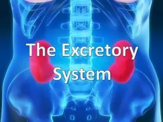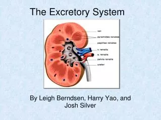
The Excretory System
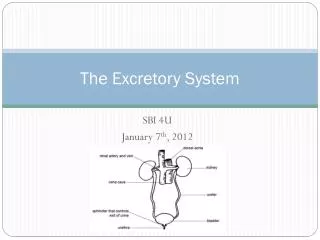
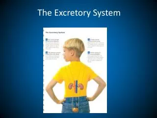
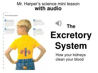
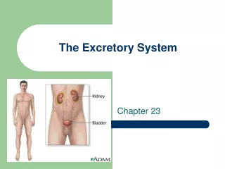
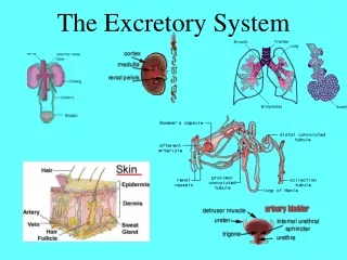
The Excretory System. Excretion- removal of waste produced during body functions. Occurs through: Intestine - digestive wastes, salts Skin (sweat glands)- water, electrolytes Lungs - carbon dioxide, water Kidneys - toxins, water, N cmpds, electrolytes. Urinary System Functions.

The Excretory System
E N D
Presentation Transcript
Excretion- removal of waste produced during body functions Occurs through: • Intestine- digestive wastes, salts • Skin (sweat glands)- water, electrolytes • Lungs- carbon dioxide, water • Kidneys- toxins, water, N cmpds, electrolytes
Urinary System Functions • maintain water concentration in blood • maintain concentration of ions like Na & K • form urine • influence rate of secretion of hormones like ADH • alter pH (acid- base balance) Why bother with all of these?
Gross Anatomy- KIDNEY • lie in retroperitoneal position • Fat cushion holds it in position • medial surface with concave hilus Not that type….
Gross Anatomy- KIDNEY • Cortex- outer and lighter • Medulla- inner and darker 1. Cortex region of kidney 2. Medulla region of kidney
Gross Anatomy- KIDNEY 6. Most of the medulla is made up of RENAL PYRAMIDS with a base facing outward and papilla facing the hilus
Gross Anatomy- KIDNEY 7. Cortical tissue dips into the medulla between the pyramids, forming RENAL COLUMNS
Gross Anatomy- KIDNEY 8. Each renal papilla juts into a cup-like CALYX • Urine leaving the renal papilla collects here before leaving the body
Gross Anatomy- KIDNEY 9. The calyces join to form the renal pelvis. It narrows as it exits the hilum to become the ureter.
10. BLOOD VESSELS Renal artery brings ¼ of all blood to kidney/min. Branches into Interlobar arteries- extend toward the cortex Changes names Arcuate arteries- base of pyramids Changes names Interlobular arteries- afferent arterioles that branch into the glomerulus where blood is filtered
10. BLOOD VESSELS (continued) Efferent arterioles Peritubular capillaries (vasa recta) Interlobular veinArcuate veinInterlobar veinrenal vein Blood flows into But, I am not going to test you on this stuff!
Macroscopic Kidneys A. Capsule & hilus B. Renal sinus 1. renal pelvis 2. major calyces 3. minor calyces C. Renal medulla 1. renal pyramids a. papilla 2. Renal column D. Renal cortex
B. Gross Anatomy- URETER • 28 cm long • Allows urine to travel from kidney to urinary bladder • 3 layers of tissue: • Mucous lining • Smooth muscle middle • DWF outer layer
C. URINARY BLADDER • behind symphysis pubis • mostly smooth muscle aka detrusor muscle lined with transitional epithelium • 3 openings: 2 from ureters and one into the urethra • Has valve to prevent backflow into kidney • Functions • urine reservoir • aided by urethra, expels urine from body
Male Urethra Female Urethra
Gross Anatomy- URETHRA • 3 cm in females; 20 cm in males • Male urethra (URINE) passes through prostate gland where it is joined by 2 ejacuatory ducts (SEMEN) then travels through penis and ends at the urinary meatus at the tip of the penis. • In females, completely separate from vagina
4. Micturition- urination • Voluntary relaxation of external sphincter muscle of bladder • Detrusor muscle contracts • Parasympathetic nerve control • Incontinence
Microscopic Structure of the NEPHRON • Filtering unit of kidney • Process blood plasma • Form urine • 1.25 million per kidney • Looks like a funnel with a long, winding stem
Components 1. renal corpuscle 2. PCT 3. loop of Henle 4. DCT 5. Collecting tubule & duct NEPHRON
The Nephron • The nephron is the functional unit of the kidney, responsible for the actual purification and filtration of the blood. • About one million nephrons are in the cortex of each kidney.
The NEPHRON RENAL CORPUSCLE- in the cortex • Bowman’s capsule • Cup-shaped mouth of nephron • Glomerulus • capillaries in BC • Pores (fenestrations) • Basement membrane
Microscopic Structure of the NEPHRON PROXIMAL TUBULE- in cortex • Closest to BC (“proximal”) • Aka PCT (proximal convoluted tubule) • Brush border (microvilli) face lumen- increase surface area
cortex medulla The NEPHRON LOOP OF HENLE (LOH) • Renal tubule beyond the PCT • Descending limb (thin) • Sharp turn • Ascending limb (thick) • Dips into medulla
THE NEPHRON DISTAL TUBULE • Aka DCT (distal convoluted tubule) • Beyond LOH (“distal”) • Juxtaglomerular apparatus
THE NEPHRON COLLECTING DUCT • Straight tubule joined by distal tubules of several nephrons • Fuse to form papillary ducts which deliver urine to the calyces
Overview of KIDNEY FUNCTION 1. FILTRATION • Occurs in glomerulus • Dependent on Glomerular Filtration Rate (GFR) • Filter water and solutes from blood into renal tubule • Glucose • Amino acids • Nitrogen wastes
KIDNEY FUNCTION-Filtration • FILTRATION • What’s left in the blood? • Blood cells • Most plasma proteins • What causes it? • Pressure gradient (high to low) • Related to blood pressure
Filtration GFR is directly dependent on blood pressure.a. If GFR (BP) is too high, filtrate flows too fast and substances are NOT reabsorbed urine flow increases water is lost blood volume drops blood pressure drops. b. If GFR (BP) is too low, filtrate flows too slow and substancesare retained too much urine flow decreases water is preserved blood volume increases blood pressure increases.
Overview of KIDNEY FUNCTION 2. REABSORPTION • Occurs in mostly in PCT and little in LOH, DCT, CD • Put good things in the renal tubule back into the blood (peritubular capillaries) • Water • Electrolytes • Nutrients
Overview of KIDNEY FUNCTION • REABSORPTION • Healthy kidneys reabsorb • Glucose (if not…) • Amino acids • Sodium • Water
Overview of KIDNEY FUNCTION • REABSORPTION • Substances that are NOT reabsorbed fully • Things that lack carriers • Things that are not lipid soluble • Things that are too large • Examples: urea, creatinine, uric acid
Overview of KIDNEY FUNCTION • REABSORPTION • ADH causes the distal and collecting tubules to become more permeable to water • This allows hypertonic urine to be formed
Overview of KIDNEY FUNCTION • SECRETION • PCT mostly • Reabsorption in reverse • Movement of small molecules out of the peritubular blood and into the tubule for excretion • Including K, H, urea, ammonia • Dispose certain drugs • Helps control blood pH
Review Questions • Do you know the names of the structures? • What is GFR? What regulates it? • Why is reabsorption important? • Where is the only place glucose is reabsorbed? • Where does ADH act? What does it do?
Making Urine • Choose your water solution (Normal or Dehydrated) • Fill the cup 2/3 of the way with that solution • Add ½ dropper of Urea + vitamins • Add ½ dropper of acid • Check pH with pH paper • If you desire, pimp your urine with 1 dropper of the following:
Answer the following questions on a piece of paper and turn it in? • Did you drink the urine? • What type did you make? • How did it taste? • Did it taste like you expected? • If you did not drink it, why not? (please provide at least three reasons)
URINE COMPOSITION • Water- 95% • Other substances- 5% • Nitrogen wastes • Electrolytes • Toxins • Pigments • Hormones • Abnormal stuff like blood, glucose, casts, calculi
URINE COMPOSITION • Characteristics • Color • Compounds • Slight odor • 4.6-8.0 pH (fresh is acidic) • 1.001- 1.035 specific gravity
FLUID, ELECTROLYTE, and ACID-BASE BALANCE • FLUID • Water accounts for 50-60% total weight (why less in obese people?) • 37% of this is ECF • 63% of this is ICF
FLUID, ELECTROLYTE, and ACID-BASE BALANCE • Mechanisms to maintain fluid balance • Volumes of ICP, ECF, plasma, and total volume of water relatively constant • Adjust output (urine volume) to intake • Adjust fluid intake (liquids we drink, water in food we eat, water formed by catabolism)
Anatomy of Micturition & Incontinence • Detrusor muscle with an External and Internal sphincter • Normal capacity 300-600cc • First urge to void 150-300cc • CNS control • Pons - facilitates • Cerebral cortex - inhibits • Harmonal effects - estrogen
Treatment Options • Reduce amount and timing of fluid intake • Avoid bladder stimulants (caffeine) • Use diuretics judiciously (not before bed) • Reduce physical barriers to toilet (use bedside commode) 1