The Excretory System
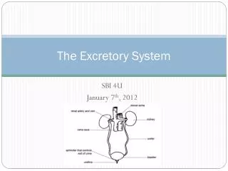
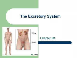
The Excretory System. By Leigh Berndsen, Harry Yao, and Josh Silver. The Urinary system. It is made of two kidneys, ureters, bladder, and Urethra. (6) The Kidneys produce urine. The urine passes though the urerter into the urinary bladder.

The Excretory System
E N D
Presentation Transcript
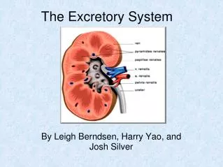
The Excretory System By Leigh Berndsen, Harry Yao, and Josh Silver
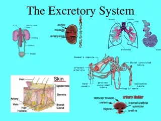
The Urinary system It is made of two kidneys, ureters, bladder, and Urethra. (6) The Kidneys produce urine. The urine passes though the urerter into the urinary bladder. It is stored in the bladder, and excreted though the urethra. The kidneys remove wastes from cellular respiration and regulate the blood concentrations (2)
Kidneys The kidneys are located against the muscles of the back just below the diaphragm, and are 10 cm long. (1) The kidney is made of three parts the cortex, medulla (inner and outer), and the pelvis. (1) The blood is filtered in the cortex, the medulla is made of collecting ducts, and the pelvis is a cavity connected to the ureter. (1,6) The collecting ducts carry the filtrate to the pelvis. (1)
Nephrons Filters the waste from the blood. Each kidney has 1.25 million nephrons. (1) Some nephrons are in the cortex, but the majority are in the medulla. Part of a nephron is a glomerulus, which is a group of capillaries. (1) Bowman’s capsule is a double walled surrounding of the glomerulus. (1) The blood in the glomerulus is filtered into the Bowman’s capsule. The filtrate exits the Bowman's capsule through the renal tubule. (3) The center section of the renal tubule is the loop of Henle, which goes to the medulla Slight reabsorbtion happens into the loop of Henle Cortical nephrons have reduced loops of Henle and jutamedullary nephrons have long loops which extends to the inner medulla. (1)
Blood in the Kidneys Blood enters through the renal arteries and exits through the renal veins. (1,6) Nephrons have arteriole, which carry the blood from the renal arteries to the nephron. (3) Prior to leaving the Bowman's capsule the capillaries join into an arteriole. The Bowman's capsule receves filtrate from the blood. These arterioles subdivide into into a second capillary network around the renal tubule. (1) Those capillaries merge into a venule. The golmerulus is a ball of capillaries from the Bowman's capsule. Afferent artiole is a renal artery that supplies blood to the nephrons. (1) Efferent artiole is when the capillaries converge and leave the capsule. (1) After the Efferent artiole, the network of capillaries subdivide into the peritubular capillaries. (1)
Renal Tubules There are three main section to this tubule: Proximal convoluted tubule, the loop of Henle, and the distal convoluted tubule. The filtrate is emptied into the collecting ducts from the renal tubules.
Urine formation The formation of urine occurs in the nephrons during two processes. Those processes are filtration and reabsorption. Filtration is a process of removing the wastes from the blood. Useful substances can be removed form the blood. (6) Reabsorption is when the useful substances/nutrients reenter the blood. (6)
Filtration Filtration takes place in the glomeruli and Bowman’s capsules. The blood in the glomerulus is under pressure. The pressure causes the filtrate which is water, urea, glucose, amino acids, and salts though the permeable walls of the glomerulus into the Bowman's capsule. (1) Blood cell and proteins are two big to pass though the walls of glomerulus Kidneys form around 180 liters of filtrate in a day Only about 1-1.5 liters of urine are produced, this is because if all the filtrate is excreted, the body will lose too much water. (3)
Reabsorption Reabsorption occurs in the renal tubule. Reabsorption reduces volume of the filtrate, and resubmit important molecules into the blood. (3) When the filtrate goes though the renal tubes of the nephron, almost all the water, glucose, amino acids and salts are reabsorbed. They enter though the capillaries around the tubules. The reabsorption of water is essential to mammals, due to homeostasis. While osmosis occurs, glucose, amino acids, and salts use active transport and ATP is supplied by the mitochondria in the renal tubules. The tubules have microvilli which increases the surface area in order to absorb. (1)
Reabsorption Crystallization in the urine is called a kidney stone Kidney threshold level, is when the concentration is too high the excess nutrients can not be absorbed. (1) The excess substances that were not reabsorbed remains in the urine and is excreted from the body. Urine flows from the tubules to the collecting ducts, then kidneys, then the ureters to the bladder, which is disposed though the urethra. (6)
Transport in the renal tubule Proximal convoluted tubule (cortex): Plays a role in homeostasis by altering volume and composition of filtrate through reaborbtion and secretion Regulates the pH throughout body fluids by secreting hydrogen ions (1) Nutrients such as glucose, amino acids, and potassium are actively transported into the interstitial fluid and then blood Microvilli on epithical cells compose the brush border allow for reabsorbtion of nutrients such as NaCl and water (1) Both active and passive transportation occur (1) Water enters the capillaries through osmosis because the solution in hypotonic compared to the cell
Descending Limb of Loop of Henle Epithelium is permeable to water and not permeable to salt and other small solutes (6) The interstitial fluid surrounding the tubule is hyperosmotic compared to the filtrate Water departs filtrate through osmosis (passive transport) (1)
Ascending limp of loop of Henle Epithelia relatively impermeable to water Thin segment: (1) permeable to NaCl NaCl diffuses out of tubule and into interstitial fluid via passive transport allows for high osmolarity in kidney Thick segment: (1) loses NaCl to interstitial fluid through active transport without giving up water filtrate is more diluted as it moves up cortex
Distal Convoluted Tubule It is a region of controlled reabsorption and secretion.(1) Contributes to pH regulation by quantitative secretion of H+ and reabsorption of bicarbonate (buffer in blood and interstitial fluid).(1) It controls the K+ and Na+ for homeostasis .(1) In order to control K+, it controls the the amount secreted into the filtrate.(1) As for Na+, it does so though reabsorption.(1)
Collecting Duct The collecting ducts carries the filtrate back in the direction of the medulla and renal pelvis. (1) The epithelium of duct is permeable to H2O, not salt. (1) The filtrate becomes more concentrated because of the loss of water though the process of osmosis (because of a hypertonic fluid outside the duct).(1) Passive transport The bottom of the duct epithelium is permeable to urea (which will be explained in the next slide), thus using passive transport.(1) Due to the bodies need to keep homeostasis when a substance has a high concentration some of the urea diffuses from the duct.(1)
Nitrogenous wastes Due to the metabolism of the body, nitrogenous wastes are formed. They generally takes the form of ammonia (NH3) and are extremely toxic to the body therefore creating a need for the body to expel it. The human body does this though a detoxification process which results in a type of waste called Urea. This is more efficient then just ammonia extraction via water (which it is extremely soluble to) because it allows a conservation of water and therefore helps the human body to retain homeostasis. (1) Kidney has high osmolarity
Brief clip describing the process of urine creation • Nephron Clip (3)
1. Campbell, Neil A. Biology. Redwood City, CA: Benjamin/Cummings, 1993. Print. 2. Google Picture. Web. 25 Oct. 2011. <http://www.google.com/imgres?q=the+excretory+system&um=1&hl=en&sa=N&rls=com.microsoft:en-us:IE-SearchBox&rlz=1I7ADFA_enUS369&biw=1004&bih=522&tbm=isch&tbnid=PHQYIn8kKwdxdM:&imgrefurl=http://www.can-do.com/uci/lessons98/BodyBuilder.html&docid=M3bXtEuOKHeGOM&imgurl=http://www.can-do.com/uci/lessons98/kidn.gif&w=398&h=427&ei=0BynTt7jJILf0QHmpfSMDg&zoom=1&iact=rc&dur=188&sig=116682043020997188527&pag e=2&tbnh=138&tbnw=129&start=10&ndsp=10&ved=1t:429,r:0,s:10&tx=57&ty=54>. 3. "Function of a Nephron." youtube. N.p., 4 Nov. 2006. Web. 25 Oct. 2011. <.http://www.youtube.com/watch?v=glu0dzK4dbU >. 4. Cartoon. BMJ. N.p., n.d. Web. 25 Oct. 2011. <http://www.bmj.com/content/319/ 7201/41.full>.5. "Nephron." Cartoon. Kidney and Nephron. N.p., n.d. Web. 25 Oct. 2011. <http://www.google.com/ imgres?q=nephrons&um=1&hl=en&client=safari&sa=N&rls=en&tbm=isch&tbnid=VTVYkd-qsYd iiM:&imgrefurl=http://oracle3927.tripod.com/ nephron.htm&docid=NbaJBK-FuVyraM&imgurl=http://oracle3927.tripod.com/images/ Nephron.jpg&w=293&h=299&ei=LzunToqUEcrj0QG-mrW9Dg&zoom=1&iact=rc&dur=274&sig=1075 Bibliogrpahy
Bibliography (cont.) 5. "Putting the Yellow in Your Urine." Cartoon. NewScientist. N.p., n.d. Web. 25 Oct. 2011. <http://www.google.com/ imgres?q=urine&um=1&hl=en&client=safari&sa=N&rls=en&biw=1042&bih=769&tbm=isch&tbn id=qOb7VCi9m2zhYM:&imgrefurl=http://scienceblogs.com/startswithabang/2009/04/ putting_the_yellow_in_your_uri.php&docid=LJOAlSh4cob3-M&imgurl=http://scienceblog s.com/startswithabang/upload/2009/04/putting_the_yellow_in_your_uri/ urine-contents.jpg&w=352&h=467&ei=KT-nTuCKGM-QswbUmZS_DQ&zoom=1&iact=rc&dur=253&s ig=107582530499020372110&page=1&tbnh=119&tbnw=90&start=0&ndsp=23&ved=1t:429,r:2,s :0&tx=10&ty=61>. 6. Starr, Cecie, and Ralph Taggart. Biology: the Unity and Diversity of Life. Pacific Grove, CA: Brooks/Cole, 2001. Print.