Chapter 10: Tissue Response to Injury
651 likes | 1.28k Vues
Chapter 10: Tissue Response to Injury. Inflammatory Response. Acute Inflammation Short onset and duration Change in hemodynamics, production of exudate, granular leukocytes Chronic Inflammation Long onset and duration Presence of non-granular leukocytes and extensive scar tissue.

Chapter 10: Tissue Response to Injury
E N D
Presentation Transcript
Inflammatory Response • Acute Inflammation • Short onset and duration • Change in hemodynamics, production of exudate, granular leukocytes • Chronic Inflammation • Long onset and duration • Presence of non-granular leukocytes and extensive scar tissue
Cardinal Signs of Inflammation • Rubor (redness) • Tumor (swelling) • Color (heat) • Dolor (pain) • Functio laesa (loss of function)
Phases of the Inflammatory Response (3 separate phases) • 1. Inflammatory response phase • 2. Fibroblastic repair phase • 3. Maturation and remodeling phase
Phase I • Healing begins immediately • Injury results in altered metabolism and liberation of various materials • Initial reaction by leukocytes and phagocytic cells • Goal • Protect • Localize • Decrease injurious agents • Prepare for healing and repair
First hour • Vasoconstriction and coagulation occur to seal blood vessels and chemical mediators are released • Immediately followed by vasodilation or blood vessel • Second hour • Vasodilation decreases blood flow, increased blood viscosity resulting in edema (swelling) • Chemical mediators • Released to alter membrane permeability, dilatory responses, margination, and phagocytic activity
Second hour (continued) • Exudate increases (high concentration of RBC’s) due to increased vessel permeability • Permeability changes generally occur in capillary and venules • Margination occurs causing leukocytes to fill the area and line endothelial walls • Through diapedesis and chemotaxis leukocytes move to injured area
Clot Formation • Platelets adhere to exposed collagen leading to formation of plug (clot) • Clots obstruct lymphatic fluid drainage and aid in localizing injury • Requires conversion of fibrinogen to fibrin • Initial stage: thromboplastin is formed • Second stage: Prothrombin is converted to thrombin due to interaction with thromboplastin • Third stage: thrombin changes from soluble fibrinogen to insoluble fibrin coagulating into a network localizing the injury
Chronic Inflammation • Occurs when acute inflammatory response does not eliminate injuring agent • Tissue not restored to normal physiologic state • Involves replacement of leukocytes with macrophages, lymphocytes and plasma cells • Cause for shift from acute to chronic is unknown • Typically associated with overuse, overload, cumulative microtrauma
Phase II: Fibroblastic Repair Phase • Scar formation through 3 phases • Resolution (little tissue damage and normal restoration) • Restoration (if resolution is delayed) • Regeneration (replacement of tissue by same tissue) • Referred to as fibroplasia • Complaints of pain and tenderness gradually subside during this period
Scar formation • Formation of delicate connective tissue (granulation tissue) • Consists of fibroblasts extracellular matrix • Develop collagen, elastin, ground substance, proteoglycans, glycosaminoglycans • With proliferation of collagen scar tensile strength increases • # of fibroblasts gradually diminishes • Normal sequence = minimal scarring • Persistent inflammation = extended fibroplasia
Phase III: Maturation & Remodeling • Long-term process • Realignment of collagen relative to applied tensile forces • Continued breakdown and synthesis of collagen = increased strength • Tissue will gradually assume normal appearance • May require several years to complete
Role of Progressive Mobilization • Initially must maintain some immobilization in order to allow for initial healing • As healing moves into repair phase controlled activity should be added • Work towards regaining normal flexibility and strength • Protective bracing should also be incorporated • During remodeling aggressive ROM and strength exercises should be incorporated • Facilitates remodeling and realignment • Must be aware of pain and other clinical signs – may be too much too soon
Extent of injury Edema Hemorrhage Poor Vascular Supply Separation of Tissue Muscle Spasm Atrophy Corticosteroids Keloids and Hypertrophic Scars Infection Humidity, Climate, Oxygen Tension Health, Age, and Nutrition Factors That Impede Healing
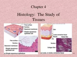
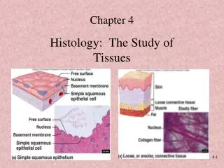
Soft Tissue Healing • Cell structure/function • All organisms composed of cells • Properties of soft tissue derived from structure and function of cells • Cells consist of nucleus surrounded by cytoplasm and encapsulated by phospholipid cell membrane • Nucleus contains chromosomes (DNA) • Functional elements of cells (organelles) include mitochondria, ribosomes, endoplasmic reticulum, Golgi apparatus & centrioles
Tissues of the Body • Bone - not classified as soft tissue • 4 types of soft tissue • Epithelial tissue • Skin, vessel & organ linings • Connective tissue • Tendons, ligaments, cartilage, fat, blood, and bone • Muscle tissue • Skeletal, smooth, cardiac muscle • Nerve tissue • Brain, spinal cord & nerves
Soft Tissue Adaptations • Metaplasia - transformation of tissue from one type to another that is not normal for that tissue • Dysplasia - abnormal development of tissue • Hyperplasia- excessive proliferation of normal cells in normal tissue arrangement • Atrophy- a decrease in the size of tissue due to cell death and re-absorption or decreased cell proliferation • Hypertrophy - an increase in the size of tissue without necessarily changing the number of cells
Cartilage Healing • Limited capacity to heal • Little or no direct blood supply • Chrondrocyte and matrix disruption result in variable healing • Articular cartilage that fails to clot and has no perichondrium heals very slowly • If area involves subchondral bone (enhanced blood supply) granulation tissue is present and healing proceeds normally
Ligament Healing • Follows similar healing course as other vascular tissues • Proper care will result in acute, repair, and remodeling phases in same time required by other vascular tissues • Repair phase will involve random laying down of collagen which, as scar forms, will mature and realign in reaction to joint stresses and strain • Full healing may require 12 months
Skeletal Muscle Healing • Initial bleeding followed by proliferation of ground substance and fibroblast • Myoblastic cells form = regeneration of new myofibrils • Collagen will mature and orient along lines of tension • Healing could last 6-8 weeks depending on muscle injured
Tendon Healing • Requires dense fibrous union of separated ends • Abundance of collagen is required for good tensile strength • Too much = fibrosis – may interfere with gliding • Initially injured tendon will adhere to surrounding tissues (week 2) • Week 3 – tendon will gradually separate • Tissue not strong enough until weeks 4-5
Nerve Healing • Cannot regenerate after injury • Regeneration can take place within a nerve fiber • Proximity of injury to nerve cell makes regeneration more difficult • For regeneration, optimal environment is required • Rate of healing occurs at 3-4 mm per day • Injured central nervous system nerves do not heal as well as peripheral nerves
Modifying Soft-Tissue Healing • Varying issues exist for all soft tissues relative to healing (cartilage, muscle, nerves) • Blood supply and nutrients is necessary for all healing • Healing in older athletes or those with poor diets may take longer • Certain organic disorders (blood conditions) may slow or inhibit the healing process
Management Concepts • Drug utilization • Anitprostaglandin agents used to combat inflammation • Non-steroidal anti-inflammatory agents (NSAID’s) • Medications will work to decrease vasodilatation and capillary permeability
Therapeutic Modalities • Thermal agents are utilized • Heat stimulates acute inflammation (but works as a depressant in chronic conditions) • Cold is utilized as an inhibitor • Electrical modalities • Treatment of inflammation • Ultrasound, microwave, electrical stimulation (includes transcutaneous electrical muscle stimulation and electrical muscle stimulation)
Therapeutic Exercise • Major aim involves pain free movement, full strength, power, and full extensibility of associated muscles • Immobilization, while sometimes necessary, can have a negative impact on an injury • Adverse biochemical changes can occur in collagen • Early mobilization (that is controlled) may enhance healing
Bone Healing • Follows same three phases of soft tissue healing • Less complex process • Acute fractures have 5 stages • Hematoma formation • Cellular proliferation • Callus formation • Ossification • Remodeling
Hematoma Formation • Trauma to the periosteum and surrounding soft tissue occurs due to the initial bone trauma • During the first 48 hours a hematoma within the medullary cavity and the surrounding tissue develops • Blood supply is disrupted by clotting vessels and cellular debris
Soft callus is a random network of woven bone • Osteoblasts fill the internal and external calluses to immobilize the site • Calluses are formed by bone fragments that bridge the fracture gap • The internal callus creates a rigid immobilization early
Hard callus formation occurs after 3-4 weeks and lasts 3-4 months • Hard callus is a gradual connection of bone filaments to the woven bone • Less than ideal immobilization produces a cartilagenous union instead of a bony union
Ossification is complete when bone has been laid down and the excess callus has been resorbed by osteoclasts • Bone continually adapts to applied stresses • Balance between osteoblast and osteoclast activity • Time required is dependent on various factors • Severity and site of fracture • Age and extent of trauma • Time will range from 3-8 weeks
Acute Fracture Management • Must be appropriately immobilized, until X-rays reveal the presence of a hard callus • Fractures can limit participation for weeks or months • A clinician must be certain that the following areas do not interfere with healing • Poor blood supply • Poor immobilization • Infection
Poor blood supply • Bone may die and union/healing will not occur (avascular necrosis) • Common sites include: • Head of femur, navicular of the wrist, talus, and isolated bone fragments • Relatively rare in healthy, young athletes except in navicular of the wrist • Poor immobilization • Result of poor casting allowing for motion between bone parts • May prevent proper union or result in bony deformity
Infection • May interfere with normal healing, particularly with compound fractures • Severe streptococcal and staphylococcal infections • Modern antibiotics has reduced the risk of infections • Closed fractures are not immune to infections within the body or blood • If soft tissue alters bone positioning, surgery may be required to ensure proper union
Healing of Stress Fractures • Result of cyclic forces, axial compression or tension from muscle pulling • Electrical potential of bone changes relative to stress (compression, tension, or torsional) • Constant stress axially or through muscle activity can impact bone resorption, leading to microfracture
If osteoclastic activity is not in balance with oesteoblastic activity bone becomes more susceptible to fractures • To treat stress fractures a balance between osteoblast and osteoclast activity must be restored • Early recognition is necessary to prevent complete cortical fractures • Decreased activity and elimination of factors causing excess stress will be necessary to allow for appropriate bone remodeling
Pain • Major indicator of injury • Pain is individual and subjective • Factors involved in pain • Anatomical structures • Physiological reactions • Psychological, social, cultural and cognitive factors
Pain Categories • Pain sources • Fast versus slow pain • Acute versus chronic • Projected or referred pain
Pain sources • Cutaneous, deep somatic, visceral and psychogenic • Cutaneous pain is sharp, bright and burning with fast and slow onset • Deep somatic pain originates in tendons, muscles, joints, periosteum and blood vessels • Visceral pain begins in organs and is diffused at first and may become localized • Psychogenic pain is felt by the individual but is emotional rather than physical
Fast versus Slow Pain • Fast pain localized and carried through A-delta axons • Slow pain is perceived as aching, throbbing, or burning (transmitted through C fibers) • Acute versus Chronic Pain • Acute pain is less than six months in duration • Chronic pain last longer than six months • Chronic pain classified by IASP as pain continuing beyond normal healing time
Referred Pain • Pain which occurs away from actual site of injury/irritation • Unique to each individual and case • May elicit motor and/or sensory response • A-alpha fibers are sensitive to pressure and can produce paresthesia • Three types of referred pain include: myofascial, sclerotomic, and dermatomic
Myofascial Pain • Trigger points or small hyperirritable areas within muscle resulting in bombardment of CNS • Acute and chronic pain can be associated with myofascial points • Often described as fibrositis, myositis, myalgia, myofasciitis and muscular strain • Active points cause obvious complaint • Trigger points do not follow patterns • Trigger point area referred to as reference zone which may or may not be proximal to the point of irritation
Sclerotomic and dermatomic pain • Deep pain with slow or fast characteristics • May originate from sclerotomic, myotomic or dermatomic nerve irritation/injury • Sclerotomic pain • Transmitted by C fibers causing deep aching and poorly localized pain • Can be projected to multiple areas of brain causing depression, anxiety, fear or anger • Autonomic changes may result (vasomotor control, BP and sweating
Dermatomic pain (irritation of A-delta fibers) is sharp and localized • Projects to the thalamus and cortex directly
Nociception • Pain receptors -free nerve endings sensitive to extreme mechanical, thermal and chemical energy • Located in meninges, periosteum, skin, teeth, and some organs • Pain information transmitted to spinal cord via myelinated C fibers and A delta fibers • Nociceptor stimulation results in release of substance P
Signal travels along afferent nerves to the spinal cord • A delta fiber (fast) transmit information to the thalamus concerning location of pain and perception of pain being sharp, bright or stabbing • C fibers (slower conduction velocity) deal with diffused, dull, aching and unpleasant pain • C fibers signal also passed to limbic cortex providing emotional component to pain