Download

1 / 97
1.04k likes | 1.77k Vues
Chapter 6: Osseous Tissue and Bone Structure. The Skeletal System. Skeletal system includes: bones of the skeleton cartilages, ligaments, and other connective tissues that stabilize the bones. Skeletal System. Functions: 1 . Support : framework & structure of body

E N D
The Skeletal System • Skeletal system includes: • bones of the skeleton • cartilages, ligaments, and other connective tissues that stabilize the bones
Skeletal System Functions: 1. Support: framework & structure of body 2. Storageof minerals and lipids Minerals: calcium and phosphate - for osmotic regulation, enzyme function, nerve impulses Yellow marrow: triglycerides 3. Blood cell production: all formed elements - red marrow: stem cells hematopiesis 4. Protection: surround soft tissues 5. Leverage for movement: - levers upon which skeletal muscles act
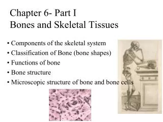
Classification of Bones • Bone are identified by: • shape • internal tissues • bone markings SHAPE: • Long bones • Flat bones • Sutural bones • Irregular bones • Short bones • Sesamoid bones
Shape of Bones • Long Bones: • Longer than wide, consist of shaft and 2 ends • e.g. bones of appendages • Short Bones: • Approx. equal in all dimensions • e.g. carpals, tarsals • Flat Bones: • Thin, 2 parallel surfaces • e.g. skull, sternum, ribs, scapula Figure 6–1a
Shape of Bones • Irregular Bones: • Complex shapes • E.g. vertebrae, os coxa • Sesamoid Bones: • Seed shaped, form in tendon • E.g. patella, total number can vary • Sutural Bones: - Extra bones in sutures of skull
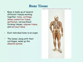
Bone Structure • A bone is an organ consisting of many tissue types: • Osseous, nervous, cartilage, fibrous CT, blood, etc. All bones consist of 2 types of bone tissue • Compact bone: - solid, dense bone, makes up surfaces and shafts • Spongy Bone/Cancellous bone: - meshy, makes up interior of bones, houses red marrow in spaces
Bone Markings • Bones are not flat on the surface: • Have projections, depressions, and holes for muscle attachment, blood & nerve supply • Depressions or grooves: • along bone surface • Projections: • where tendons and ligaments attach • at articulations with other bones • Tunnels: • where blood and nerves enter bone
Bone Markings Table 6–1 (2 of 2)
Long Bones Structure • Diaphysis: - Hollow shaft of compact bone • Medullary (marrow) cavity: • Center of diaphysis, contains yellow marrow • Triglycerides for energy reserve • Epiphysis: • Expanded end of bone, surface of compact bone • Center filled with spongy bone with red marrow in spaces • Produces blood cells Figure 6–2a
Long Bones Structure • Epiphyseal line or plate: • Cartilage that marks connection of diaphysis with epiphysis • Line: adults, narrow (aka metaphysis) • Plate: thick, allows growth during childhood • Periosteum: • 2 layer covering around outside of bone: • Outer Fibrous Layer • Inner Cellular Layer • Endosteum: • Cellular layers, covers all inside surfaces
Articular Cartilage: • Hyaline cartilage on end where bone contacts another, no periosteum or perichondrium Joint/Articulation: - connection between two bones, surrounded by CT capsule, lined with synovial membrane Joint cavity filled with synovial fluid to reduce friction on articular cartilage
Flat Bone Structure • Thin layer of spongy bone with red marrow between two layers of compact bone • Covered by periosteum and endosteum • Site of most hematopoiesis • Production of blood cells and cell fragments that are suspended in plasma (RBC, WBC, and platelets
Characteristics of Bone Tissue • Periosteum: • covers outer surfaces of bones • consist of outer fibrous and inner cellular layers • Endosteum: • Inner, cellular layer of periosteum
Bone Histology • Bone = osseous tissue, supporting CT • Consists of specialized cells in a matrix of fibers and ground substance • Characteristics of bone: • Dense matrix packed with calcium salts • Osteocytes in lacunae • Canaliculi for exchange of nutrients and waste • Two layer periosteum, covers bone except at articular surfaces
Bone Histology • Matrix = 98% of bone tissue • 1/3 = osteoid; organic part: • Collagen fibers + ground substance • Tough and flexible • 2/3 = densely packed crystals of hydroxyapatite (calcium salts, mostly calcium phosphate) • Hard but brittle • Cells = only 2% of bone • Osteocytes • Osteoblasts • Osteoprogenitor cells • Osteoclasts
Cells located in Bones • Osteocytes = mature bone cells -no cell division -located in lacunae between layers of matrix called lamellae -canaliculi link lacunae to each other and blood supply -osteocytes linked to each other via gap junctions on cell projections in canaliculi: - allow exchange of nutrients and wastes -Function 1. To maintain protein and mineral content of matrix 2. Can also participate in bone repair: -become stem cell like when broken free of lacuna
Canaliculi Osteocytes in lacunae PERIOSTEUM Fibrous layer Blood vessels Cellular layer Central canal Matrix LM X 362
Cells located in Bones • Osteoblasts - Immature bone cells • Perform osteogenesis: • Formation of new bone matrix • Produce osteoid • Organic components of matrix that is not yet calcified to form bone • Promote deposit of calcium salts which spontaneously form hydroxyapatite • Once enclosed in lacuna by matrix, osteoblast differentiates into osteocyte and no longer produces new matrix
Cells located in Bones 3. Osteoprogenitor Cells – mesenchymal cells - bone stem cell that produces daughters - daughters become osteoblasts for repair and growth - located in endosteum and inner periosteum
Cells located in Bones 4. Osteoclasts - large, multinuclear - derived from monocytes (macrophages) - perform osteolysis = - digest and dissolve bone matrix - release minerals: 1. For use in blood or 2. Recycling during bone remodeling
Osteoprogenitor cell Canaliculi Osteocyte Matrix Osteoid Osteoblast Matrix Osteoclast Matrix Marrow cavity Osteocyte: Mature bone cell that maintains the bone matrix Osteoblast: Immature bone cell that secretes organic components of matrix Osteoprogenitor cell: Stem cell whose divisions produce osteoblasts Osteoclast: Multinucleate cell that secretes acids and enzymes to dissolve bone matrix Cells located in Bones
Homeostasis • Bone building (by osteocytes) and bone recycling (by osteoclasts) must balance: • more breakdown than building, bones become weak • exercise causes osteocytes to build bone
How would the strength of a bone be affected if the ratio of collagen to hydroxyapatite increased? Strength increases, flexibility increases. Strength increases, flexibility decreases. Strength decreases, flexibility. decreases. Strength decreases, flexibility increases.
If the activity of osteoclasts exceeds the activity of osteoblasts in a bone, how will the mass of the bone be affected? stable mass, but re-positioned matrix mass will not be affected more mass less mass
Structure of Compact Bone • Consists of osteons: • Parallel to surface • Each osteon is around a central canal: • Contains blood vessels and nerves • Perforating canals perpendicular to osteons act to connect the osteons • Osteon is built of layers of matrix secreted by osteoblasts • Each layer = concentric lamella • Osteocytes are located in lacunae between lamellae • Ostocytes are connected to neighboring cells and central canal via canaliculi
Structure of Compact Bone • Interstitial lamellae fill spaces between osteons • Circumferiential lamellae run perimeter inside and out in contact with: • endosteum and periosteum • Compact bone is designed to receive stress from one direction • Very strong parallel to osteons • Weak perpendicular to osteons
Compact Bone Figure 6–5
Structure of Spongy Bone • Lamellae = meshwork called trabeculae (no osteons) • Red marrow fills spaces around trabeculae • Osteocytes in lacunae are linked by canaliculi • No direct blood supply (no central canals) • Nutrients diffuse into canaliculi in trabeculae from red marrow • Spongy bone make up: • low stress bones • Areas of bone where stress comes from multiple directions • Provide light weigh strength
Bone Marrow • Red Marrow: • Located in space between trabeculae • Has blood vessels • Forms red blood cells • Supplies nutrients to osteocytes • Yellow Marrow: • In some bones, spongy bone holds yellow bone marrow: • is yellow because it stores fat
Periosteum and Endosteum • Compact bone is covered with membrane: • periosteum on the outside • endosteum on the inside
Periosteum • Fibrous outer layer: - Dense irregular CT • Cellular Inner layer: • Osteoprogenitor cells Functions: • Isolate bone from surrounding tissues • Site for attachment for tendons and ligaments • Route for nerves and blood vessels to enter bone • Participates in bone growth and repair
Endosteum • Thin cellular layer • Lines medullary cavity, central canals, and covers trabeculae • Consists of: • osteoblasts, osteoprogenitor cells, and osteoclasts • Cells become active during bone growth and repair
Endosteum Figure 6–8b
Bone Growth • Begins 6-8 weeks post fertilization • Continues through puberty (18-25 y) • Osteogenesis = ossification = formation of bone • Not calcification • Hardening of matrix or cytoplasm with calcium • Can happen to many tissues • Two types of Ossification: • Intramembranous: forms flat bones • Endochondrial: forms long bones
Bone Development • Human bones grow until about age 25 • Osteogenesis: • bone formation • Ossification: Deposition of calcium salts • the process of replacing other tissues with bone
The difference between intramembranous ossification and endochondral ossification.
Intramembranous Ossification • Bone develops from mesenchyme or fibrous CT in deep layers of dermis • Also called dermal ossification: • because it occurs in the dermis • produces dermal bones such as mandible and clavicle • Produces skull bones • There are 4 main steps in intramembranous ossification
Intramembranous Ossification: Step 1 • Ossification center appears in the fibrous CT membrane • Mesenchymal cells aggregate • Differentiate into osteoblasts • Begin ossification at the ossification center
Intramembranous Ossification: Step 2 • Bone matrix (osteoid) is secreted within the fibrous membrane • Osteoblasts begin to secrete osteoid, which is mineralized within a few days • Trapped osteoblasts become osteocytes
Intramembranous Ossification: Step 3 • Woven bone and periosteum form • Accumulating osteoid is laid down between embryonic blood vessels, which form a random network • Vascularized mesenchyme condenses on the external face of the woven bone and becomes periosteum around spongy bone
Intramembranous Ossification: Step 4 • Bone collar of compact bone forms and red marrow appears • Trabeculae just deep to the periosteum thickens, forming a woven bone collar that is later replaced with mature lamellar bone • Spongy bone, consisting of distinct trabeculae, persists internally and its vascular tissue becomes red marrow
Endochondral Ossification • Ossifies bones that originate as hyaline cartilage • Most bones originate as hyaline cartilage • Cartilage grows by interstitial and appositional growth • Cartilage is slowly replaced from the inside out