Download
1 / 40
400 likes | 632 Vues
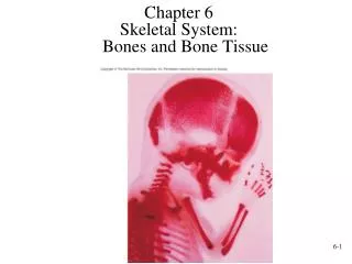
Skeletal System: Bones and Bone Tissue. Chapter 6. Functions of the Skeletal System. Support . B one is hard and rigid; cartilage is flexible yet strong. Cartilage in nose, external ear, thoracic cage and trachea. Ligaments- bone to bone

E N D
Skeletal System:Bones and Bone Tissue Chapter 6
Functions of the Skeletal System • Support. Bone is hard and rigid; cartilage is flexible yet strong. Cartilage in nose, external ear, thoracic cage and trachea. Ligaments- bone to bone • Protection. Skull around brain; ribs, sternum, vertebrae protect organs of thoracic cavity • Movement. Produced by muscles on bones, via tendons. Ligaments allow some movement between bones but prevent excessive movement (joint: formed where two or more bones come together------allows movement between bones) • Storage. Ca and P. Stored then released as needed. Fat stored in marrow cavities • Blood cell production. Bone marrow that gives rise to blood cells and platelets
Components of Skeletal System • Bone • Cartilage: three types • Hyaline • Fibrocartilage • Elastic • Tendons and ligaments (Tendons: muscle to bone Ligament: bone to bone)
Hyaline Cartilage • Consists of specialized cells that produce matrix • Chondroblasts: form matrix • Chondrocytes: surrounded by matrix; are lacunae • Matrix. Collagen fibers for strength, proteoglycans for resiliency • Perichondrium. Double-layered C.T. sheath. Covers cartilage except at articulations • Inner. More delicate, has fewer fibers, contains chondroblasts • Outer. Blood vessels and nerves penetrate. No blood vessels in cartilage itself • Articular cartilage. Covers bones at joints; has no perichondrium • Growth • Appositional. New chondrocytes and new matrix at the periphery (chondroblasts in perichondrium lay down new matrix & new chondrocytes) • Interstitial. Chondrocytes within the tissue divide and add more matrix between the cells.
Bone Histology • Bone matrix. Like reinforced concrete. Steel bars is collagen fibers, cement is hydroxyapatite • Organic: collagen and proteoglycans (35%) • Inorganic: hydroxyapatite. CaPO4 crystals (65%) • If mineral removed, bone is too bendable • If collagen removed, bone is too brittle
Bone Histology • Bone cells • Osteoblasts • Osteocytes • Osteoclasts • Stem cells or osteochondral progenitor cells • Woven bone: collagen fibers randomly oriented • Lamellar bone: mature bone in sheets • Cancellous bone (spongy): contain trabeculae -less bone matrix & more space • Compact bone: dense -more bone matrix & less space
Bone Cells • Osteoblasts • Formation of bone through ossification or osteogenesis. Collagen produced by E.R. and golgi. Released by exocytosis. Precursors of hydroxyapetite stored in vesicles, then released by exocytosis. • Ossification: formation of bone by osteoblasts. It occurs by appositional growth on surface of previously existing bone. Osteoblasts communicate through gap junctions. Bone matrix produced by osteoblasts covers the older bone & surrounds osteoblasts results in new layer of bone.
Bone Cells • Osteocytes. Mature bone cells once surrounded by matrix. They are relatively inactive, but can make small amounts of matrix to maintain it. • Lacunae: spaces occupied by osteocyte cell body • Canaliculi: canals occupied by osteocyte cell processes • Nutrients diffuse through tiny amount of liquid surrounding cell and filling lacunae and canaliculi. Then can transfer nutrients from one cell to the next through gap junctions.
Bone Cells • Osteoclasts. Responsible for resorption of bone (breakdown of bone) • Ruffled border (projections): where cell membrane borders bone and resorption is taking place. • H ions pumped across membrane, acid forms, eats away bone. • Release enzymes that digest the bone. • Derived from monocytes (which are formed from stem cells in red bone marrow) • Multinucleated • Stem Cells. Mesenchyme(Osteochondral Progenitor Cells) become chondroblasts or osteoblasts. (Remember: osteocytes derived from osteoblasts)
Woven and Lamellar Bone • Woven bone. Collagen fibers randomly oriented. - first formed during fetal development or repair of fracture - after formation, osteoclasts break down woven bone & osteoblasts build new matrix---------------this process called remodeling • Remodeling • Removing old bone and adding new • Woven bone is remodeled into lamellar bone • Lamellar bone • Mature bone in sheets called lamellae. Fibers are oriented in one direction in each layer, but in different directions in different layers for strength.
Cancellous (Spongy) Bone • Trabeculae: interconnecting rods or plates of bone. Like scaffolding. • Spaces filled with marrow & blood vessels. • Covered with endosteum. • Oriented along stress lines • Trabeculae consist of several lamellae (thin sheets) w/ osteocytes located in lacunae between them
Compact Bone • Central or Haversian canals: parallel to long axis • Lamellae: thin sheets/layers concentric (circles), circumferential (perimeter, periphery), interstitial (between osteon) • Osteon or Haversian system: central canal (also called haversian canals------contains blood vessels that run parallel to long axis of bone), associated concentric lamellae and osteocytes • Perforating or Volkmann’s canal: perpendicular to long axis. Both perforating and central canals contain blood vessels. Central canals receive blood vessels from perforating canals & nutrients in blood enter central canals, pass into canaliculi, move through osteocytes to osteocytes (by gap junctions). Waste removed in reverse direction.
Bone Shapes • Long • Ex. Upper and lower limbs • Short • Ex. Carpals and tarsals • Flat • Ex. Ribs, sternum, skull, scapulae • Irregular • Ex. Vertebrae, facial
Structure of a Long Bone • Diaphysis • Shaft • Compact bone • Epiphysis • End of the bone • Cancellous bone • Epiphyseal plate: growth plate (separates epiphysis from diaphysis) • Hyaline cartilage; present until growth stops • Epiphyseal line: bone stops growing in length (epiphyseal plate becomes epiphyseal line) • Medullary cavity (space):In children medullary cavity is red marrow, gradually changes to yellow in limb bones and skull (except for epiphyses of long bones). Rest of skeleton is red.
Structure of a Long Bone, cont. • Periosteum • Outer is fibrous (contains blood vessels & nerves) • Inner is single layer of bone cells including osteoblasts, osteoclasts and osteochondral progenitor cells • Tendons & ligaments attach to bone & become continuous with fibers of periosteum. • Sharpey’s fibers: some collagen fibers of tendons or ligaments penetrate the periosteum and into the bone. Strengthen attachment of tendon or ligaments to bone. • Endosteum. Similar to periosteum, but more cellular. Lines all internal surfaces of all cavities including spaces in cancellous bone.
Structure of Flat, Short, and Irregular Bones • Flat Bones • No diaphyses, epiphyses • Sandwich of cancellous between compact bone • Short and Irregular Bone • Compact bone that surrounds cancellous bone center; similar to structure of epiphyses of long bones (bec. have processes that possess epiphyseal growth plates • No diaphyses and not elongated • Some flat and irregular bones of skull have sinuses (air filled spaces) lined by mucous membranes.
Bone Development(during fetal development) • Intramembranous ossification • Takes place in connective tissue membrane • Endochondral ossification • Takes place in cartilage • Both methods of ossification • Produce woven bone that is then remodeled • After remodeling, formation cannot be distinguished as one or other
Intramembranous Ossification (8th week – 2 years of development) • Takes place in connective tissue membrane formed from embryonic mesenchyme • Forms many skull bones, part of mandible, diaphyses of clavicles • When remodeled, indistinguishable from endochondral bone. • Centers of ossification: locations in membrane where ossification begins (centers of ossification expand outwards to form a bone by gradually ossifying the membrane) • Fontanels: large membrane-covered spaces between developing skull bones; unossified (bones eventually grow together & all fontanels have closed by 2 years of age)
Endochondral Ossification • Bones of the base of the skull, part of the mandible, epiphyses of the clavicles, and most of remaining bones of skeletal system • Cartilage formation begins at end of fourth week of development • Some ossification beginning at about week eight; some does not begin until 18-20 years of age
Growth in Bone Length • Growth in length occurs at the epiphyseal plate • Involves the formation of new cartilage by • Interstitial cartilage growth • Appositional growth on the surface of the cartilage • Closure of epiphyseal plate: epiphyseal plate is ossified becoming the epiphyseal line. Between 12 and 25 years of age • Articular cartilage: does not ossify, and persists through life • Appositional growth only • Interstitial growth cannot occur because matrix is solid • Occurs on old bone and/or on cartilage surface ex: trabeculae grow in size by the deposition of new bone matrix by osteoblasts onto the surface of the trabeculae.
Growth at Articular Cartilage • Increases size of bones with no epiphyses: e.g., short bones • Chondrocytes near the surface of the articular cartilage similar to those in zone of resting cartilage • The process of growth in articular cartilage is similar to that occurring in the epiphyseal plate, except that the chondrocyte columns are not as obvious. • When epiphyses reach their full size, the growth of cartilage & its replacement by bone cease. • However, articular cartilage persists throughout life & does not become ossified as does epiphyseal plate.
Factors Affecting Bone Growth • Size and shape of a bone determined genetically but can be modified and influenced by nutrition and hormones • Nutrition • Lack of calcium, protein and other nutrients during growth and development can cause bones to be small • Vitamin D • Necessary for absorption of calcium from intestines • Can be eaten or manufactured in the body • Rickets: lack of vitamin D during childhood (bowed bones) • Osteomalacia: lack of vitamin D during adulthood leading to softening of bones • Vitamin C • Necessary for collagen synthesis by osteoblasts • Scurvy: deficiency of vitamin C (causes ulceration & hemorrhage in body) • Lack of vitamin C also causes wounds not to heal (because requires collagen synthesis), teeth to fall out (because ligaments that hold them in place break down)
Factors Affecting Bone Growth (cont.) • Hormones • Growth hormone from anterior pituitary. Stimulates interstitial cartilage growth and appositional bone growth • Thyroid hormone required for growth of all tissues • Sex hormones such as estrogen and testosterone • Cause growth at puberty, but also cause closure of the epiphyseal plates and the cessation of growth
Bone Remodeling • Converts woven bone into lamellar bone • Caused by migration of Basic Multicellular Units (BMU) • Groups of osteoclasts and osteoblasts that remodel bones • Involved in bone growth, changes in bone shape, adjustments in bone due to stress, bone repair, and Ca ion regulation • Relative thickness of bone changes as bone grows. Bone constantly removed by osteoclasts and new bone formed by osteoblasts. • Formation of new osteons in compact bone • Osteoclasts enter the osteon from blood in the central canal and internally remove lamellae. Osteoblasts replace bone • Osteoclasts remove bone from the exterior and the bone is rebuilt
Bone Repair • Hematoma formation. Localized mass of blood released from blood vessels (damaged from fracture) but confined within an organ or space. Clot formation (consists of fibrous proteins that stop the bleeding). • Callus formation. Callus: mass of tissue that forms at a fracture site and connects the broken ends of the bone. - Internal- blood vessels grow into clot in hematoma (several days after fracture). • Macrophages clean up debris, osteoclasts break down dead tissue, fibroblasts produce collagen and granulation tissue. • Chondroblasts from osteochondral progenitor cells of periosteum and endosteum produce cartilage within the collagen. • Osteoblasts invade. New bone is formed. - External- collar around opposing ends. Periosteal osteochondral progenitor cells osteoblasts and chondroblasts. Bone/cartilage collar stabilizes two pieces.
Bone Repair, cont. • Callusossification. Callus replaced by woven, cancellous bone through endochondral ossification. • Bone remodeling. Replacement of cancellous bone and damaged material by compact bone. Sculpting of site by osteoclasts
Calcium Homeostasis • Bone is major storage site for calcium • The level of calcium in the blood depends upon movement of calcium into or out of bone. • Calcium enters bone when osteoblasts create new bone; calcium leaves bone when osteoclasts break down bone • Two hormones control blood calcium levels- parathyroid hormone (produced by parathyroid gland----blood calcium decreases then secretion of PTH increases which increases # of osteoclasts) and calcitonin ( produced by thyroid gland----decreases osteoclast activity
Effects of Aging on Skeletal System • Bone matrix decreases. More brittle due to lack of collagen; but also less hydroxyapatite. • Bone mass decreases. Highest around 30. Men denser due to testosterone and greater weight. African Americans and Hispanics have higher bone masses than Caucasians and Asians. Rate of bone loss increases 10 fold after menopause. Cancellous bone lost first, then compact. • Increased bone fractures • Bone loss causes deformity, loss of height, pain, stiffness • Stooped posture • Loss of teeth
Bone Fractures • Open (compound)- bone break with open wound. Bone may be sticking out of wound. • Closed (simple)- Skin not perforated. • Incomplete- doesn’t extend across the bone. Complete- does • Greenstick: incomplete fracture that occurs on the convex side of the curve of a bone • Hairline: incomplete where two sections of bone do not separate. Common in skull fractures • Comminuted fractures: complete with break into more than two pieces
Bone Fractures, cont. • Impacted fractures: one fragment is driven into the cancellous portion of the other fragment. • Classified on basis of direction of fracture • Linear • Transverse • Spiral • Oblique • Dentate: rough, toothed, broken ends • Stellate radiating out from a central point.


