
Chapter 7
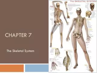
Chapter 7. The Skeletal System. Introduction. Individual bones are the organs of the skeleton system Bone contains very active tissues. Bone Structure. Bone structure reflects its function Parts of a long bone Epiphyses: Located at each end Covered by articular cartilage

Chapter 7
E N D
Presentation Transcript
Chapter 7 The Skeletal System
Introduction • Individual bones are the organs of the skeleton system • Bone contains very active tissues
Bone Structure • Bone structure reflects its function • Parts of a long bone • Epiphyses: • Located at each end • Covered by articular cartilage • Articulates (forms joint) with other bones • Diaphysis: • Shaft of bone
Bone Structure Cont. • Parts of a long bone cont. • Periosteum • Covers bone • Mostly found on diaphysis • Compact Bone • Provides strength and resistance to bending • Spongy Bone • Provides strength where needed and reduces weight of bone • Medulllary Cavity • In diaphysis, it’s the area filled with marrow
Microscopic Structure • Osteocytes • Mature bone cells • Lacunae • Bony chambers • Processes • Bony projections that provide a site for ligament and tendon attachment
Microscopic Structure Cont. • Osteons • Found in compact bone • Are cemented together to form cylinder shaped units (Haversian System)
Microscopic Structure Cont. • Osteonic Canals (Haversian Canals) • Contain blood vessels that nourish the cells of the osteons • Spongy bone is nourished by diffusion from the surface of the thin bony plates.
Bone Development and Growth • Intramembranous Bones • Develop from layers of connective tissues • Osteoblasts within the membranous layers form bone tissue
Bone Growth and Development Cont. • Endochondral Bones • Developed first as hyaline cartilage to be later replaced by bone tissue • Primary Ossification (creation of new bone tissue) center appears in the diaphysis, Secondary Oss. In epiphysis.
Bone Growth and Development Cont. • Epiphyseal Disk • Division between primary and secondary ossification centers • Responsible for lengthening bone • Bone continues to lengthen until disks ossify. • Growth in thickness is due to intramembranous ossification beneath periosteum • If Epi. Disk is damaged before ossified, it can cause premature growth ending.
Bone Function • 5 Functions • 1. Support • 2. Protection • Bones shape and form body structures • Bones support and protect softer, underlying tissues
Bone Function Cont. • 3. Body Movement • Bones and muscles function together as levers • A lever consists of a rod, pivot (fulcrum), a movable weight, and a force that supplies energy.
Bone Function Cont. • 4. Blood Cell Formation • Hematopoiesis (development of blood cells) • At different ages, hematopoiesis occurs in the yolk sac, liver and spleen, and red bone marrow. • Red Bone marrow produces red blood cells (RBC’s), white blood cells (WBC’s) and blood platelets • Yellow Marrow stores fat
Bone Function Cont. • 5. Storage of Inorganic Salts • Intercellular material of bone tissue contains large quantities of calcium phosphate. • When blood calcium is low, osteoclasts break down bone, when blood calcium is high, osteoblasts build bone. • Bone stores small amounts of magnesium, sodium, potassium and carbonate ions.
Skeletal Organization p. 142-149 • Axial Portion • Skull • Hyoid bone • Vertebral column • Thoracic cage • Appendicular Portion • Pectoral girdle • Upper limbs • Pelvic girdle • Lower limbs
Skull • Consists of 22 bones • 8 cranial • 13 facial • 1 mandible
Cranium • Encloses and protects the brain • Some bones contain air filled sinuses • Include • Frontal • Parietal • Occipital • Temporal • Sphenoid • Ethmoid
Facial Skeleton • Provides the basic shape of face and attachments for muscles • Includes: • Maxillary • Palatine • Zygomatic • Lacriminal • Nasal • Vomer • Inferior nasal conchae • mandible
Infantile Skull • Fontanels separate incompletely developed bones • Proportions of the infantile skull are different from those of an adult skull.
Table 7.2 Skeletal Structure Terms • Condyle • Definition: A rounded process that usually articulates with another bone • Example: Occipital condyle of occipital bone • Crest • Defintion: A narrow, ridge-like projection • Example: Iliac crest of ilium • Epicondyle • Defintion: A projection situated above a condyle • Example: Medial epicondyle of humerus • Facet • Definition: A small, nearly flat surface • Example: Rib-facet of thoracic vertebra • Fontanel • Definition: A soft spot in the skull where membranes cover the spaces between bones • ExampleAnterior fontanel between frontal and parietal bones
Table 7.2 Skeletal Structure Terms Cont. • Foramen • Definition: An opening through a bone that usually is a passageway for blood vessels, nerves, or ligaments • Example: Foramen magnum of occipital bone • Fossa • Definition: A relatively deep pit or depression • Example: Olecranonfossaofhumerus • Fovea • Definition: A tiny pit or depression • Example: Fovea capitis of femur • Head • Definition: An enlargement of the end of a bone • Example: Head of humerus • Meatus • Definition: A tube-like passageway within a bone • Example: External auditory meatus of ear
Table 7.2 Skeletal Structure Terms Cont. • Process • Definition: A prominent projection on a bone • Example: Mastoid process of temporal bone • Sinus • Definition: A cavity within a bone • Example: Frontal sinus of frontal bone • Spine • Definition: A thorn-like projection • Example: Spine of scapula • Suture • Definition: An interlocking line of union between bones • Example: Lambdoidal suture between occipital and parietal bones • Trochanter • Definition: A relatively large process • Example: Greater trochanter of femur
Table 7.2 Skeletal Structure Terms Cont. • Tubercle • Definition: A small, knob-like process • Example: Greater tubercle of humerus • Tuberosity • Definition: A knob-like process usually larger than a tubercle • Example: Radial Tuberosity of radius
Vertebral Column p. 149-155 • Extends from skull to the pelvis • Protects the spinal cord • Composed of vertebrae • Separated by intervertebral disks • Has four curvatures
Typical Vertebra • Consists of: • A body • A bony arch (surrounds spinal cord) • Contains notches on upper and lower surfaces that provide intervertebral foramina through which spinal nerves pass.
Cervical Vertebrae (7) • Atlas • 1st vertebra-supports and balances head • Axis • 2nd vertebra-contains dens that provide intervertebral foramina through which spinal nerves pass • Contains Transverse Process which bears the transverse foramina
Thoracic Vertebrae (12) • Larger than cervical • Contains facets on the sides that articulate with the ribs
Lumbar Vertebrae (5) • Vertebral Bodies are large and strong • Support more body weight than other vertebrae
Saccrum • Is a triangular structure formed of five fused vertebrae • Vertebral foramina form the sacral canal
Coccyx • Composed of four fused vertebrae • Forms the lowest part of vertebral column • Your TAILBONE!
Thoracic Cage • Includes: • Ribs • Thoracic vertebra • Sternum • Costal cartilages • Supports the shoulder girdle and arms • Protects visceral organs • Functions in breathing
Ribs • 12 Pairs are attached to the thoracic vertebrae • True Ribs join sternum directly by costal cartilages • False Ribs join indirectly or not at all • Typical rib has a shaft, head, and tubercles that articulate with the vertebrae.
Sternum • Consists of three parts: • Manubrium • Body • Xiphoid process • Articulates with clavicles
Pectoral Girdle P. 155 • Composed of: • Two clavicles • Two scapula • Forms an incomplete ring that supports the upper limbs and provides attachment for muscles
Clavicles • Rod-like bones located between the manubrium and scapulae • Hold the shoulders in place and provide attachment for muscles
Scapula • Broad, triangular bones • Articulate with the humerus of each upper limb and provide attachment for muscles
Upper Limbs • Provide framework and attachment for muscles • Function in levers that move the limb and its parts • Contains: • Humerus • Radius • Ulna • Hand
Humerus • Extends from the scapula to the elbow • Articulates with the radius and ulna at elbow
Radius • Located on the thumb side of the forearm between the elbow and wrist • Articulates with humerus, ulna and wrist
Ulna • Longer than radius • Overlaps the humerusposteriorly • Articulates with the radius laterally and with a disk of fibrocartilage inferiorly
Carpals (8) • Consists of 8 bones: • Lunate • Hamate • Triquetrum • Pisiform • Scaphoid • Capitate • Trapezoid • Trapezium • Articulates with the radius and fibrocartilaginous disk on ulnar side
Metacarpals (5) • Consists of 5 bones: • Metacarpals 1-5 • Cylindrical, rounded distal ends that form the knuckles • Articulate proximally with carpals and distaly with phalanges
Phalanges (14) • Finger Bones • Each finger has three phalanges (digits) • A proximal, a middle and a distal phalanx (digit) • The Thumb has two-it lacks a middle phalanx


