Download
1 / 42
420 likes | 841 Vues
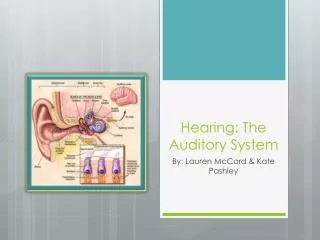
Auditory Sensation (Hearing) L13. Faisal I. Mohammed, MD, PhD. Objectives. Define decibel (intensity) and Hz (frequency) Describe the ossicular system and explain its function Follow up sound transmission up to the cochlea Outline the structure of cochlea, and the organ of Corti

E N D
Auditory Sensation (Hearing)L13 Faisal I. Mohammed, MD, PhD University of Jordan
Objectives • Define decibel (intensity) and Hz (frequency) • Describe the ossicular system and explain its function • Follow up sound transmission up to the cochlea • Outline the structure of cochlea, and the organ of Corti • Describe the mechanism of sound transduction • Follow up the auditory pathway to the cerebral cortex • Describe auditory abnormalities (types of deafness) University of Jordan
Frequency of sound wave:Audible sound wave pure tones20 – 20,000 Hz Speed of sound is 335 m/sec in Air University of Jordan
Decibel: a measure of sound intensity • Decibel (dB) = 10 log I/IR • I = intensity of sound, IR= reference intensity • Acoustic intensity is proportional to the square of sound pressure level • Sound pressure is more conveniently measured than sound intensity • Sound pressure level (SPL) unit is decibel • SPL (dB) = 20 log P/PR • P= the sound pressure in N/m2 (N=Newton, m = meter) • PR= reference pressure (either 0.0002 dynes/cm2, the absolute threshold for human hearing and equal 20 micropascal, or 1 dyne/ cm2 ) University of Jordan
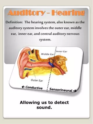
The Tympanic Membrane and the Ossicular System • Tympanic membrane functions to transmit vibrations in the air to the cochlea • Amplifies the signal because the area of the tympanic membrane is 17 times larger than the oval window (55 sq. mm Vs. 3.2 sq. mm) • Tympanic membrane connected to the ossicles • malleus • incus • Stapes • Ossicular system works as a lever system and amplifies the sound 1.3 time • Total amplification is 22 times (17x1.3) called Impedance Matching (match the resistance of sound wave movement in fluid vs. the resistance in air) University of Jordan
Components of the Auditory System University of Jordan
Attenuation of Sound by Muscle Contraction • Two muscles attach to the ossicles • Stapedius (supplied by facial Nn VII) • tensor tympani (supplied by Trigeminal Nn V) • A loud noise initiates reflex contraction after 40 - 80 milliseconds • Attenuates vibration going to cochlea (Attenuation reflex) • Serves to protect cochlea and damps low frequency sounds i.e., your own voice University of Jordan
Cochlea • system of three coiled tubes separated by membranes into the scala tympani, scala media, scala vestibuli • sound waves cause back and forth movement of the tympanic membrane which moves the stapes back and forth • this causes displacement of fluid in the cochlea and induces vibration in the basilar membrane University of Jordan
Structure of the Human Cochlea University of Jordan
BasilarMembrane • contains about 30,000 fibers which project from the bony center of the cochlea, the modiolus • fibers are stiff reed-like structures fixed to the modiolus and embedded in the loose basilar membrane • because they are stiff and free at one end they can vibrate like a musical reed • the length of the fibers increase and the diameter of the fibers decrease from base to the helicotrema, overall stiffness decreases 100 times, high frequency resonance occurs near base, low near apex University of Jordan
Structural Components of the Cochlea University of Jordan
Organ of Corti • receptor organ that generates nerve impulses • lies on the surface of the basilar membrane, contains rows of cells with stereocilia called hair cells • the tectorial membrane lies above the stereocilia of the hair cells • movement of the basilar membrane causes the stereocilia of the hair cells to shear back and forth against the tectorial membrane University of Jordan
The Organ of Corti University of Jordan
movement of the basilar membrane causes the stereocilia of the hair cells to shear back and forth against the tectorial membrane. University of Jordan
Stapes vibrating in oval window Stapes vibrating in oval window Stapes vibrating in oval window Stapes vibrating in oval window Stapes vibrating in oval window Stapes vibrating in oval window Stapes vibrating in oval window Stapes vibrating in oval window Stapes vibrating in oval window Incus Incus Incus Incus Incus Incus Incus Incus Incus Malleus Malleus Malleus Malleus Malleus Malleus Malleus Malleus Malleus Cochlea Cochlea Cochlea Cochlea Cochlea Cochlea Cochlea Cochlea Cochlea Helicotrema Helicotrema Helicotrema Helicotrema Helicotrema Helicotrema Helicotrema Helicotrema Helicotrema Sound waves Sound waves Sound waves Sound waves Sound waves Sound waves Sound waves Sound waves Sound waves Perilymph Perilymph Perilymph Perilymph Perilymph Perilymph Perilymph Perilymph Perilymph Scala tympani Scala tympani Scala tympani Scala tympani Scala tympani Scala tympani Scala tympani Scala tympani Scala tympani 3 3 3 3 3 3 3 8 8 7 7 7 4 4 4 4 4 4 Scala vestibuli Scala vestibuli Scala vestibuli Scala vestibuli Scala vestibuli Scala vestibuli Scala vestibuli Scala vestibuli Scala vestibuli 5 5 5 5 5 6 6 6 6 Basilar membrane Basilar membrane Basilar membrane Basilar membrane Basilar membrane Basilar membrane Basilar membrane Basilar membrane Basilar membrane 1 1 1 1 1 1 1 1 1 2 2 2 2 2 2 2 2 9 External auditory canal External auditory canal External auditory canal External auditory canal External auditory canal External auditory canal External auditory canal External auditory canal External auditory canal 8 8 Spiral organ (organ of Corti) Spiral organ (organ of Corti) Spiral organ (organ of Corti) Spiral organ (organ of Corti) Spiral organ (organ of Corti) Spiral organ (organ of Corti) Spiral organ (organ of Corti) Spiral organ (organ of Corti) Spiral organ (organ of Corti) Tectorial membrane Tectorial membrane Tectorial membrane Tectorial membrane Tectorial membrane Tectorial membrane Tectorial membrane Tectorial membrane Tectorial membrane Vestibular membrane Vestibular membrane Vestibular membrane Vestibular membrane Vestibular membrane Vestibular membrane Vestibular membrane Vestibular membrane Vestibular membrane Cochlear duct (contains endolymph) Cochlear duct (contains endolymph) Cochlear duct (contains endolymph) Cochlear duct (contains endolymph) Cochlear duct (contains endolymph) Cochlear duct (contains endolymph) Cochlear duct (contains endolymph) Cochlear duct (contains endolymph) Cochlear duct (contains endolymph) Tympanic membrane Tympanic membrane Tympanic membrane Tympanic membrane Tympanic membrane Tympanic membrane Tympanic membrane Tympanic membrane Tympanic membrane Secondary tympanic membrane vibrating in round window Secondary tympanic membrane vibrating in round window Secondary tympanic membrane vibrating in round window Secondary tympanic membrane vibrating in round window Secondary tympanic membrane vibrating in round window Secondary tympanic membrane vibrating in round window Secondary tympanic membrane vibrating in round window Secondary tympanic membrane vibrating in round window Secondary tympanic membrane vibrating in round window Middle ear Middle ear Middle ear Middle ear Middle ear Middle ear Middle ear Middle ear Middle ear Auditory tube Auditory tube Auditory tube Auditory tube Auditory tube Auditory tube Auditory tube Auditory tube Auditory tube University of Jordan
Nerve Impulse Origination • The stereocilia, when bent in one direction cause the hair cells to depolarize, and when bent in the opposite direction hyperpolarize. • this is what begins the neural transduction of the auditory signal • Auditory signals are transmitted by the inner hair cells. • 3-4 times as many outer hair cells than inner hair cells • outer hair cells may control the sensitivity of the inner hair cells for different sound pitches University of Jordan
Determination of Sound Frequency and Amplitude (sound intensity) • Place principle determines the frequency of sound perceived. • Different frequencies of sound will cause the basilar membrane to oscillate at different positions (basilar membrane is tonotopically organized) • Position along the basilar membrane where hair cells are being stimulated determines the pitch of the sound being perceived. • Phase-locked (volley) principle at lower frequencies of sound where firing rate determines the phase of sound wave (i.e frequency of sound wave) • Amplitude is determined by how much the basilar membrane is displaced ( by frequency of impulses from the nerve fiber and the No. of nerve fibers stimulated) University of Jordan
The “Place Principle” University of Jordan
Threshold of hair cells University of Jordan
Decibel Unit of Sound • unit of sound • expressed in terms of the logarithm of their intensity • a 10 fold increase in energy is 1 bel • 0.1 bel is a decibel • 1 decibel is an increase in sound energy of 1.26 times University of Jordan
Central Auditory Pathway University of Jordan
from medial geniculate to auditory cortex from inferior colliculus to medial geniculate from superior olivary nucleus to inferior colliculus via the lateral lemniscus some fibers pass to the ipsilateral superior olivary nucleus. 2nd order neurons project through trapezoid body to the contralateral superior olivary nucleus. fibers enter dorsal and ventral cochlear nuclei of the upper part of the medulla. University of Jordan
Auditory Cortex and Association Areas - arranged by tonotopic maps - high frequency sounds at one end of map - low frequency sounds at other end - discrimination of sound patterns University of Jordan
Determining the Direction of Sound • superior olivary nucleus divided into lateral and medial nuclei • lateral nuclei detects direction by the difference in sound intensities between the two ears • medial nuclei detects direction by the time lag between acoustic signals entering the ears University of Jordan
Deafness • nerve deafness • impairment of the cochlea or the auditory nerve • conduction deafness • impairment of tympanic membrane or ossicles University of Jordan
Audiometery University of Jordan
Weber test Rinne test University of Jordan
The Internal Ear (Site of Equilibrium Receptors) University of Jordan
Thank You University of Jordan