Download
1 / 19
200 likes | 378 Vues
THE OPTICAL ABSORPTION SPECTRA OF Fe 2+ and Fe 3+ IONS IN THE BLOOD. Maksims Poļakovs Institute of Solid State Physics, University of Latvia, Kengaraga street 8, LV-1063 Riga, Latvia. The aims of work. Geometrical parameters of erythrocytes

E N D
THE OPTICAL ABSORPTION SPECTRA OF Fe2+ and Fe3+ IONS IN THE BLOOD Maksims Poļakovs Institute of Solid State Physics, University of Latvia, Kengaraga street 8, LV-1063 Riga, Latvia
The aims of work • Geometrical parameters of erythrocytes • In the present work we report results of measurements optical absorption of blood.
Used equipments • For optical absorption spectra measurements was used “Specord UV-VIS”. • For geometrical parameters of erythrocytes was used Automatic analyzer of microscopic image “MORPHQUANT”
Materials Venous blood was donated by consenting Chernobyl clean-up workersand healthy men. Blood was collected under air in glass tubes containing a small amount of heparinused as an anticoagulant. Blood without any anticoagulant wasalso testedand showed no difference with respect heparinblood.
Clinical investigations show high morbidity rate of clean-up workers compare with general population. Nowadays they represent group of chronically sick people with diseases prevalence of digestive, musculoskeletal and nervous system. Most of the Chernobyl’ clean-up workers have poli-symptomatic sicknesses that exhibit tendency to progress.
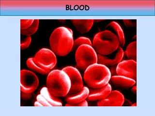
Some of clean-up workers have erythrocytosis.Erythrocytosis is defined as an excess of erythrocytes, or red blood cells (RBCs).RBCs in the blood are measured by the hematocrit (the percentage of the blood volume made up of RBCs) or by the hemoglobin
erythrocytes The diameter of a typical human erythrocyte disk is 6–8 µm, much smaller than most other human cells. A typical erythrocyte contains about 270 million hemoglobin molecules, with each carrying four heme groups. Adult humans have roughly 2–3 × 10¹³ red blood cells at any given time (women have about 4 million to 5 million erythrocytes per cubic millimeter (microliter) of blood and men about 5 million to 6 million; people living at high altitudes with low oxygen tension will have more). Red blood cells are thus much more common than the other blood particles AFM image of erythrocytes
Geometrical parametrs of erythrocytes (RBC) P - Perimeter of RBCS - Square of RBCf - Form factorof RBC (P2/S) D - The maximal size of RBCW - The minimal size of RBCExt - Eccentricity of RBC ( D/W )
Result: Slim - parametrs of erytrocytes for Chernobyl`s clean-up wokers Norma – parametrs of erytrocytes for practically heath peopele
Hemoglobin structure • Hemoglobin is nanoparticlewith sizes 6,4 x 5,5 x 5,0 nm • Composed of four globular protein subunits (tetramer) each with a heme group. • Four types of globin chains occur in pairs containing 141-146 amino acids • Responsible for cells ability to transport O2 and CO2 • When a ligandis bound to Hb, the heme ironis 6-coordinated
Heme structure of Hemoglobin Ion of iron Fe2+ is bounding up with molecule of O2 does not change valence, but transferring from high to low spin state.
Hemoglobin Fe 2+ low and high spin state Fe2+ [Ar]3d6 Dq high spin < Dq low spin eg eg t2g t2g Low spin S=0 (oxyhemoglobin) High spin S=2 (deoxyhemoglobin)
Methemoglobin Fe(II)-heme is oxidized to Fe(III)-heme to form metHb. MetHb does not bind O2. oxy-Hb met-Hb
oxyHb(Fe2+) metHb(Fe3+)
Methemoglobin Fe 3+ low and high spin state Fe3+ [Ar]3d5 Dq high spin < Dg low spin eg eg t2g t2g Low spin S=1/2 High spin S=5/2
Optical absorption spectra of oxy-hemoglobin (Fe2+ ; S=0) Absorption spectrum of Methemoglobin and Hemoglobin
CONCLUSIONS We observed that geometrical parameters of erythrocytes for healthy people differ from geometrical parameters for sick people. We observed the additional absorption bands 630-660 nm in absorption spectra of blood of Chernobyl clean–up worker who has erythrocytosis. These data shows that the blood of the Chernobyl`s clean –up workers which have erythrocytosis met hemoglobin higher normal. Perhaps the ion Fe 2+ in the-heme of hemoglobin is oxidized to the Ion Fe3+ in the heme by radiation. While estimating the health status of clean-up workers part of the revealed pathology could be explained by the higher levels of strontium and other radionucleids in the body.