Download
1 / 32
320 likes | 420 Vues
Biology Of Human Aging. Chapter 12 Urinary System. Outline. Review of the Structure and Function Kidneys / Ureters / Urinary Bladder / Urethra Age-Related Changes Kidneys / Bladder and Urethra Age-Related Dysfunctions Urinary iIncontinence Nocturia

E N D
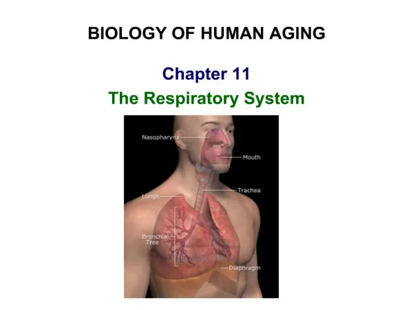
Biology Of Human Aging Chapter 12 Urinary System
Outline • Review of the Structure and Function • Kidneys / Ureters / Urinary Bladder / Urethra • Age-Related Changes • Kidneys / Bladder and Urethra • Age-Related Dysfunctions • Urinary iIncontinence • Nocturia • Dysfunctions Caused by the prostate gland • Pyelonephritis • Renal Calculi
Kidney Functions • Filter 200 liters of blood daily, allowing toxins, metabolic wastes, and excess ions to leave the body in urine • Regulate volume and chemical makeup of the blood • Maintain the proper balance between water and salts, and acids and bases • Production of rennin to help regulate blood pressure and erythropoietin to stimulate RBC production • Activation of vitamin D
Other Urinary System Organs • Urinary bladder – provides a temporary storage reservoir for urine • Paired ureters – transport urine from the kidneys to the bladder • Urethra – transports urine from the bladder out of the body
Urinary System Organs Figure 25.1a
Layers of Tissue Supporting the Kidney • Renal capsule – fibrous capsule that prevents kidney infection
Internal Anatomy • A frontal section shows three distinct regions • Cortex– the light colored, granular superficial region • Medulla – exhibits cone-shaped medullary (renal) pyramids • Pyramids are made up of parallel bundles of urine-collecting tubules • Renal pelvis – flat, funnel-shaped tube lateral to the hilus within the renal sinus
Internal Anatomy • Major calyces – large branches of the renal pelvis • Collect urine draining from papillae • Empty urine into the pelvis • Urine flows through the pelvis and ureters to the bladder
Internal Anatomy Figure 25.3b
The Nephron • Nephrons are the structural and functional units that form urine, consisting of: • Glomerulus – a tuft of capillaries associated with a renal tubule • Glomerular (Bowman’s) capsule – blind, cup-shaped end of a renal tubule that completely surrounds the glomerulus
The Nephron • Renal corpuscle – the glomerulus and its Bowman’s capsule • Podocytes (cleft cells)
Renal Tubule • Proximal convoluted tubule (PCT) –with numerous microvilli and mitochondria • Reabsorbs water and solutes from filtrate and secretes substances into it • Loop of Henle – a hairpin-shaped loop of the renal tubule • Distal convoluted tubule (DCT) – cuboidal cells without microvilli that function more in secretion than reabsorption
Renal Tubule Figure 25.4b
Capillary Beds Figure 25.5a
Capillary Beds of the Nephron • Every nephron has two capillary beds • Glomerulus • Peritubular capillaries • Each glomerulus is: • Fed by an afferent arteriole • Drained by an efferent arteriole
Capillary Beds of the Nephron • Blood pressure in the glomerulus is high because: • Arterioles are high-resistance vessels • Afferent arterioles have larger diameters than efferent arterioles
Filtration Membrane Figure 25.7a
Filtration Membrane Figure 25.7c
Mechanisms of Urine Formation • Urine formation and adjustment of blood composition involves three major processes • Glomerular filtration • Tubular reabsorption • Secretion Figure 25.8
Ureters • Slender tubes that convey urine from the kidneys to the bladder • Ureters enter the base of the bladder through the posterior wall • This closes their distal ends as bladder pressure increases and prevents backflow of urine into the ureters • Ureters actively propel urine to the bladder via response to smooth muscle stretch
Urinary Bladder • Smooth, collapsible, muscular sac that temporarily stores urine • Outlined by the openings for the ureters and the urethra • Clinically important because infections tend to persist in this region
Urinary Bladder Figure 25.18a, b
Urethra • Muscular tube that: • Drains urine from the bladder • Conveys it out of the body
Urethra • Sphincters keep the urethra closed when urine is not being passed • Internal urethral sphincter – involuntary sphincter at the bladder-urethra junction • External urethral sphincter – voluntary sphincter surrounding the urethra as it passes through the urogenital diaphragm
Age-Related Changes • Kidney • Thickening of connective tissue capsule • Decrease in thickness of cortical region • General atrophy of cells and gradual decrease in kidney weight • Decrease in kidney functioning • Increase in the incidents of abnormal glomeruli and replacement of degenerated glomeruli by connective tissue (non-permeable) • Proteinuria (presence of protein in urine) • Increased abnormalities and degeneration of renal tubules • Blood vessels: thickening of walls, loss of muscular tissue, and atherosclerotic deposits • Decline in the ability to handle large changes in acid base levels
Age-Related Changes • 2. Bladder and Urethra • Weakening and loss of elasticity of the muscles in the walls • Inability to expand or contract lower bladder capacity in elderly • Retention of residual urine (100 ml) after urination in elderly • Delay in awareness of the need to urinate • Urination in elderly may be urgent, & due to weakness of external urethral sphincter elderly may be unable to reach the lavatory in time • Urination during night more common in elderly • Weakness of the muscles of the pelvic cavity floor ineffectiveness of external urethral sphincter urine leakage (usually seen in sudden rise in pressure of bladder) cough or sneeze This is calledStress Incontinence (more common in older women)
Age-Related Dysfunctions • Urinary Incontinence • Involuntary passing of urine through the urethra • Incidents twice as high in women as in men • Due to weakness of internal & external urethral sphincters & uninhibited contractions of smooth muscles hyperactive bladder • Significant problem in older persons marketing of adult diaper-like undergarments is a profitable enterprise • Also reduction of bladder volume & delayed sensation to urinate due to muscle atrophy • Post-menopausal women: estrogen deficiency weak muscles • Involuntary urine passage is due to: • Mechanical conditions that elevate bladder pressure (cough or sneeze) • Incomplete emptying of the bladder
2. Nocturia • Excessive urination at night • Not a serious condition by itself, yet disturbs sleep • Taking drugs: bodily reactions to drugs (relaxation of sphincters or stimulation of muscles) • Not taking drugs: due to age related losses in bladder distensibility & inability of kidney to concentrate here
3. Dysfunctions caused by the Prostate Gland • Reproductive organ, yet interferes with function of urinary system • Overall atrophy of prostate with aging, yet a lot of men experience growth of prostate • Benign hyperplasia of prostate: compresses the urethra, difficulty in urination, bladder never completely emptied • Increases pressure in glomerular capsules kidney malfunction • Symptoms: Reduced force behind urine stream, frequent urination, inability to empty bladder • Treatment: surgery, transurethral resection
3. Dysfunctions caused by the Prostate Gland • Carcinoma: most frequent tumor of old men • Early stages: little or no urethral obstruction, unaware of tumor • Later stage: restrict urine flow, tumor too far advanced to be treated • Regular examination, Digital Rectal Exam (DRE), PSA test • Drugs: lengthy remissions, surgery
4. Pyelonephritis • Inflammation of the kidney • Not restricted to older persons • Acute pyelonephritis is a bacterial infection that travels to the kidney in the blood or lymphs • Causes swelling of kidney due to fluid build up • In severe cases, abscesses develop inflammation & pus • Chronic problem and causes extensive scar tissue in kidney kidney failure • Responds well to antibiotic treatment • Serious condition (uremia)
5. Renal Calculi • Kidney stones, not restricted to elderly • Most stones do not cause significant symptoms • Passage of stones from kidney, ureter, & bladder strong muscle contractions episodes of severe pain • If stone lodged in ureter obstructs urine flow kidney damage, ulceration, vulnerable to infection • Cause: Ca2+, uric acid, cystine, magnesium, ammonium phosphate • Kidney stone formation related to: Various kidney infections, high salt concentration in urine, Vit. A deficiency, tumor of parathyroid gland