Download
1 / 13
140 likes | 283 Vues
Improved Design for Fine-Needle Aspiration (FNA) of Breast Cancer Lesions. Alissa Garman Janie Goldsworthy Kristi Hinner Nick Kortan Client: Elizabeth Burnside Advisor: John Webster Midsemester Presentation March 7, 2003. Problem Statement.

E N D
Improved Design for Fine-Needle Aspiration (FNA) of Breast Cancer Lesions Alissa Garman Janie Goldsworthy Kristi Hinner Nick Kortan Client: Elizabeth Burnside Advisor: John Webster Midsemester Presentation March 7, 2003
Problem Statement GOAL:To modify the needle used during a Fine Needle Aspiration (FNA) procedure. The modification should not drastically change the FNA procedure and still increase the amount of tissue removed for cytological testing. It should also minimize the time (by collecting an adequate sample size on the first attempt) and discomfort caused to the patient during the procedure.
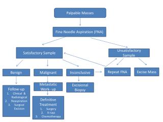
What is FNA? • Technique used to extract cells; no incision needed • Several insertions are usually required to ensure an adequate sample size • Samples are sent to pathologist to be analyzed National Breast Cancer Center, http://www.NBCC.org
Fine Needle Aspiration (FNA) A.D.A.M., http://www.adam.com
Product Design Specifications • Removes adequate number of cells for diagnosis. • Should not cause more discomfort than current FNA procedure. • Constrained to standard 20-25-gauge needle, client prefers 23-gauge.
Current Prototype • Pre-manufactured drill bit • 6” length • Fits in 23 gauge needle • Bead on shaft of drill bit to allow for ease of turning during cell collection
Tissues Tested • Fixed cat tissues (Last semester) • Mammary • Lymph • Fat • Fresh tissue • Mouse mammary tumor (cystic)
Testing Procedures (Fresh Tissue) • Old technique • No insert used • Performed one trial on the mouse tumor • New technique • Drill bit insert used • Performed one trial on the mouse tumor • Observed in saline bath under microscope • Prepared slides and sent to lab for evaluation
Sample Quantification • Old technique • Resulted in a small amount of tissue • New technique • Roughly five times as much tissue collected using drill bit
Slide Preparation • Two methods • Air-dry method - Place sample on slide and smear using a second slide - Results in two identical slides - Stain with Methanol Blue type stain • ThinPrep method - Place sample in vile of ThinPrep solution - Slide prepared using ThinPrep machine in lab - $8.00/vile
Sample Slides Benign FNA Cell Sample
Sample Slides Malignant FNA Cell Samples
Future Work • Test on more freshly sacrificed mouse tumors • Have slides evaluated by pathologist • In vivo testing of mouse tumors- will require protocol amendment • Further prototype development • Develop protocol for quantifying testing • Continued research on independent cytopathological evaluation