PLACENTA
PLACENTA. Dr. Ahmed Fathalla Ibrahim. DECIDUA. DEFINITION: It is the functional layer of endometrium in a pregnant women It includes the endometrium of fundus & body of uterus


PLACENTA
E N D
Presentation Transcript
PLACENTA Dr. Ahmed Fathalla Ibrahim
DECIDUA • DEFINITION: • It is the functional layer ofendometrium in a pregnant women • It includes the endometrium of fundus & body of uterus • The endometrium of the cervix does not form a part of decidua. The cervix is closed by mucous plug during pregnancy
DECIDUA • CAUSE OF FORMATION: • Trophoblast secretes chorionic gonadotrophins which prolong the life of corpus luteum • The corpus luteum of pregnancy continues to secrete progesterone • The increasing level of progesterone in maternal blood induces endometrial changes into decidua
DECIDUA • STEPS OF FORMATION (DECIDUAL REACTION): IMPORTANT IN DIAGNOSIS OF EARLY PREGNANCY • The endometrium becomes thicker & more vascular • The endometrial glands become full of secretion • The connective tissue cells enlarge due to accumulation of lipid & glycogen. They are called “decidual cells”
DECIDUA • PARTS OF DECIDUA: • Decidua basalis: between chorionic sac & muscle wall of uterus • Decidua capsularis: between chorionic sac & cavity of uterus • Decidua parietalis: remaining part
DECIDUA • FATE OF DECIDUA: The chorionic sac expands towards the cavity of the uterus due to the growth of fetus & enlargement of amniotic cavity: • The cavity of uterus is obliterated due to fusion of chorion & amnion • The decidua parietalis & capsularis disappear gradually • The decidua basalis persists to form a part of placenta
CHORION & CHORIONIC VILLI • DEVELOPMENT: During implantation, the trophoblast thickens at region of contact with endometrium & differentiates into: • Outer syncytiotrophoblast: a layer containing multiple nuclei in a common cytoplasm with no cell boundaries (secretes HCG) • Inner cytotrophoblast: a layer of mononucleated cells
STAGES OF CHORIONIC VILLI • PRIMARY: At the end of 2nd week,finger-like processes formed of outer syncytiotrophoblast & inner cytotrophoblast appear • SECONDARY: Early in 3rd week, extraembryonic mesoderm extends inside the villi • TERTIARY: During 3rd week, arterioles, venules & capillaries develop in the mesenchyme of villi & join umbilical vessels By the end of 3rd week, embryonic blood begins to flow slowly through capillaries in chorionic villi
CHORIONIC VILLI • Chorionic villi invade & erode endometrium, maternal blood leaks & collects in spaces between villi (intervillous spaces) • Cytotrophoblast penetrates syncytiotrophoblast & extends to form a cytotrophoblastic shell that surrounds the chorionic sac & attaches it to endometrium. It prevent further penetration of decidua by syncytiotrophoblast
TYPES OF CHORIONIC VILLI • STEM (ANCHORING) VILLI: attach the chorionic sac to decidua through cytotrophoblastic shell • BRANCH (TERMINAL) VILLI: grow from the sides of stem villi, increase surface area for exchange between fetal & maternal blood
FATE OF CHORIONIC VILLI • Until the beginning of 8th week, chorionic villi surround the whole chorionic sac, then differentiate into: • Smooth chorion (chorion laeve): villi in contact with decidua capsularis, degenerate & become avascular due to compression & disappearance of decidua capsularis • Villous chorion (chorion frondosum): largevilli in contact with decidua basalis, form part of placenta
DEVELOPMENT OF PLACENTA • FETAL PART: VILLOUSCHORION • MATERNAL PART: DECIDUA BASALIS
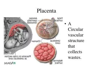
PLACENTA • MATERNAL SURFACE B) FETAL SURFACE
PLACENTA • SHAPE: circular disc • WEIGHT: about one-sixth that of fetus (500-600 gm) • DIAMETER: 15-20 cm • THICKNESS: 2-3 cm • SURFACES: • Fetal surface: smooth, transparent, covered by amnion with umbilical cord attached near its center & umbilical vessels radiating from it • Maternal surface: irregular, divided into convex areas (cotyledons)
PLACENTA • The fetal part of placenta is divided into cotyledons by projections from decidua basalis (placental septa) • Each cotyledon contains 2 or more stem villi with their branch villi • By the end of 4th month, decidua basalis is almost entirely replaced by cotyledons
PLACENTAL CIRCULATION • Poorly oxygenaed blood leaves the fetus & passes through the 2 umbilical arteries to the placenta • Umbilical arteries divide into several chorionic arteries that enter chorionic villi • An arterio-capillary-venous network is formed in chorionic villi • Exchange of materials between fetal & maternal blood (in intervillous spaces) occurs across placental membrane (barrier) • Oxygenated blood returns to fetus by umbilical vein
PLACENTAL MEMBRANE • It separates fetal from maternal blood & prevents mixing of them • It is an incomplete barrier because it only prevents large molecules to pass (heparin, bacteria), but cannot prevent passage of viruses (e.g. rubella), microorganisms (e.g. toxoplasma gondii, treponema pallidum), drugs, hormones
PLACENTAL MEMBRANE COMPONENTS: • Until 20th week: it is formed of 4 layers: • Syncytiotrophoblast • Cytotrophoblast • Extraembryonic mesoderm • Endothelium of fetal blood vessels • After 20th week: it is more permeable to meet increased demand of fetus & is formed of 2 layers : • Syncytiotrophoblast • Endothelium of fetal blood vessels
FUNCTIONS OF PLACENTA • METABOLISM: synthesis of glycogen, cholesterol & fatty acids: source of nutrition & energy for embryo or fetus during early pregnancy
FUNCTIONS OF PLACENTA • TRANSFER:transport of substances in both directions between placenta & maternal blood: • Gases:oxygen, carbon dioxide • Nutritional substances:glucose, aminoacids, vitamins • Hormones:steroid hormones (e.g. testosterone) • Electrolytes: Na+, K+, Cl- • Maternal antibodies:antibodies against diphteria, smallpox, measles
FUNCTIONS OF PLACENTA • SECRETION OF HORMONES: • Protein hormones: • Human chorionic gonadotropin (early pregnancy tests) • Human placental lactogen • Human chorionic thyrotropin • Human chorionic corticotropin • Relaxin • Steroid hormones: • Progesterone • Estrogen
ANOMALIES OF PLACENTA • ANOMALIES IN SIZE & SHAPE: normally chorionic villi persist only at site of decidual basalis: • Placenta membranacea: a large placenta due to persistence of on the entire surface of chorionic sac • Accessory placenta: a patch of chorionic villi persisted a short distance from main placenta
ANOMALIES OF PLACENTA • ANOMALIES IN POSITION: • Placenta previa: when blastocyst implants close to or overlying internal os of uterus late pregnancy bleeding • ANOMALIES IN EXTENSION: • Placenta accreta: chorionic villi extend to the myometrium • Placenta percreta: chorionic villi penetrate the whole thickness of myometrium & extend to perimetrium
BATTLEDORE PLACENTA VELAMENTOUS INSERTION OF CORD
ANOMALIES OF PLACENTA • ANOMALIES IN ATTACHMENT OF UMBILICAL CORD: • Battledore placenta: the cord is attached to the margin of placenta • Velamentous insertion of cord: the cord is attached to the membranes surrounding placenta, umbilical vessels are liable to be ruptured