Download

1 / 85
860 likes | 1.19k Vues
DISEASES OF RBCs. MAJI JOSE. DISORDERS OF RBC ANEMIA IRON DEFICENCY ANEMIA PLUMMR VINSON SYNDROME PERNICIOUS ANEMIA MEGALOBLASTIC ANEMIA APLASTIC ANEMIA SICKLE CELL ANEMIA THALASSEMIA

E N D
DISEASES OF RBCs MAJI JOSE
DISORDERS OF RBC ANEMIA IRON DEFICENCY ANEMIA PLUMMR VINSON SYNDROME PERNICIOUS ANEMIA MEGALOBLASTIC ANEMIA APLASTIC ANEMIA SICKLE CELL ANEMIA THALASSEMIA ERYTHROBLASTOSIS FETALIS POLYCYTHEMIA
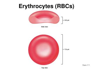
Normal range • 5.4 million/cu mm in males • 4.8 million/cu mm in females Pathological variation in number • Polycythemia- above 7mil/cu mm • Anemia- decreased
Variation in shape • Crenation Projections from the surface(Alkaline PH) • Spherocytosis Hereditary spherocytosis, autoimmune hemolytic anemia & ABO hemolytic disease of newborn. • Sickle cells or drepanocytes Sickle cell disease
Poikilocytosis • Schistocytosis Fragmentation of RBC Thalassemia, megaloblastic & Fe deficiency anemia, severe burns. • Leptocytosis Thinned RBC eg: target cells. Fe deficiency anemia, thalassemia. • Acanthocytosis Coarsely crenated RBC. Post splenectomy, chronic liver disease.
Burr cells • Cell fragments with >1 spines. • Uraemia. • Stomatocytes • Central slit like appearance. • Alcoholism.
Variation in structure • Punctatebasophilia • Basophilic granularity in RBC. • Aplastic anemia, thalassemia, myelodysplasia, infections & lead poisoning. • Howell jolly bodies • Purple nuclear remnants. • Megaloblastic anemia.
RBC Indices • RBC count Normal : 5.5 million/mm3 (males) 4.8 million/mm3 (females) • Hb Normal : 13-18g/dl (males) 11.5-16.5g/dl (females) • PCV Normal : 45ml/100ml
MCV Normal : 77-93 fl • MCH Normal : 27-32 pg • MCHC Normal : 27-32 g/dl
DISORDEROFRBC Anemia State in which the hemoglobin level in the blood is below the lower extreme of the normal range for the age & sex of the individual. PREVALENCE : F > M AGE : young age.
Etiological classification of anemia- wintrobe Loss of blood Acute post hemorrhagic anemia Chronic post hemorrhagic anemia. • Anaemia due to impaired red cell production • 1.Impaired blood production resulting from • Deficient haem synthesis - Fe def anemia • Deficient vitamins- vit B 12/folic acid. • 2.Inadequate production of mature erythrocytes • Defecency of erythroblast • - Atrophy of bone marrow: aplasia anemia. • -isolated erythoblastopenia. • Bone marrow infiltration • Leukemias • Lymphomas • Myelosclerosis • Multiple myeloma • endocrine abnormality • Myxedema • Addisionionadernalinsufficency • Thyroidism • chronic renal disease • chronic inflammatory disease • -infection • -Non infectious deasease • Cirrhosis of liver Anaemia • excessive distruction of RBC • extracorpuscular cause: • antibiotics • infection • Splenicdistruction • Drugs,chemicals • Truma of RBC • -intracorpuscular cause • Hereditary: • Disorder of glycolysis. • Abnormality in synthesis of globin. • Abnormal RBC membrane. • Erythropoiticprophyria. • Aquired : • Lead poisining
Excessive destruction of RBC Extracorpuscular cause: • antibiotics • infection • Splenicdistruction • Drugs,chemicals • Truma of RBC Intracorpuscular cause • Hereditary: • Disorder of glycolysis. • Abnormality in synthesis of globin. • Abnormal RBC membrane. • Erythropoiticprophyria. • Aquired : • Lead poisining
Anaemia due to impaired red cell production • 1.Impaired blood production resulting from • Deficient haem synthesis - Fe def anemia • Deficient vitamins- vit B 12/folic acid. • 2.Inadequate production of mature erythrocytes • Defecency of erythroblast • Atrophy of bone marrow: aplasia anemia. • isolated erythoblastopenia. • 3. Bone marrow infiltration • Leukemias • Lymphomas • Myelosclerosis • Multiple myeloma • 4. Endocrine abnormality • Myxedema • Addisionionadernalinsufficency • Thyroidism • - chronic renal disease • 5. chronic inflammatory disease • -infection • 6.Non infectious disease • Cirrhosis of liver
MORPHOLOGIC CLASSIFICATION • MACROCYTIC • Lack of erythrocyte maturing factors. • NORMOCYTIC • Hemorrhage, hemolysis, lack of blood formation & dilution of blood with fluid. • SIMPLE MICROCYTIC • Infections & inflammatory diseases. • HYPOCHROMIC MICROCYTIC • Fe deficiency.
IRON DEFECIENCY ANEMIA Most prevalent single deficiency state. Most prevalent anemia in Indians , females Etiology • Increased blood loss • Uterine – menstruation , post menopausal uterine bleeding, repeated miscarriages • Gastrointestinal – peptic ulcer, hookworm infestation, cancer, varices, ulcerative colitis, aspirin ingestion. • Renal tract – hematuria, haemoglobinuria • Nose – epistaxis
Increased requirement • Growth spurts( 1- 2.5mg /day) • Pregnancy & lactation( 1.5-2.5 mg/day) • Inadequate dietary intake • Poor economic status • Anorexia • Elderly individuals • Decreased absorption • Partial gastrectomy • Achlorhydria • Intestinal malabsorption
Clinical features • Mainly reported in females in 4th/5th decay of life • Lassitude, weakness, fatigue, • dyspnoea on exertion & palpitations • Lemon tinted pallor of skin & mucous membrane, conjunctiva . • Splenomegaly • Koilonychia and brittle nails. • Numbness and tingling of finger ,toes and bone pain.
Oral manifestations • Glossitis and stomatitis with atrophy of papillae(bald tongue). • Pallor of mucosa • Oral candidiasis, burning mouth, recurrent apthous ulcers, • angular cheilitis
Investigation • Blood examination -hypochromatic microcytic anemia. • Hb is low with RBC count 3,000,000 to4,000,000 cells/cu.mm • Bone marrow aspiration-reveals absence of stainable iron.
Biochemical findings – • Plasma ferritin- measure iron store and is a specific test. (150 – 2000ng /dl) • Plasma iron – low. (Serum iron level 80 – 180ug /dl) • Elevated total iron binding capacity (250 – 450 ug /dl) • Transferrin saturation < 16% is consistent finding • Increased soluble transferrin receptor.
Histological alterations: • Deficiency of keratinized cells. • Reduced cytoplasmic diameter of cells with paradoxical enlargement of nucleus. • Abnormal cellular maturation: Disturbed nuclear pattern, increase in nucleoli, double nuclei & karyorrhexis
DIAGNOSIS • Careful reasoned assessment of the cause. • Based on definitive investigation: serum ferritin level <30microgrm/l • bone marrow aspirates which fails to stain with perssian blue. indicated stored iron depletion.
ORAL HEALTH CONSIDERATION • Surgical procedure may result in bleeding and poor wound healing. • Hemorrhagic manipulation should be deferred for a minimum of 2wk. • Hb values below 10g pt is in risk for elective dental treatment and GA
MANAGEMENT - iron supplementation. - oral discomfort is managed by topical rinse with local anesthetics
PLUMMER – VINSON SYNDROME • SYNONYM: Paterson kelly syndrome / sideropenic dysplasia. • A rare syndrome with triad of dysphagia, iron deficiency anemia and upper esophageal web/strictures. • Age and sex: middle age women between 4th-7th decayed. • Etiology: iron deficiency anemia malnutrition autoimmune genetic disorders
CLINICAL FEATURES • Dysphagia: intermittent /progressive. • Weight loss • Symptoms of anemia • Splenomegaly • Carcinoma of upper alimentary tract also reported.
Oral manifestation • Cracked or fissured corner of mouth. • Painful tongue with atrophy of papillae • Esophageal webbing can be demonstrated radiographically
Oral health consideration • Physician consultation • Hb<8grms/dl GA is avoided MANAGEMENT • Same as iron deficiency anemia. • Early diagnosis and sooner treatment with iron supplements will resolve webs which are not substantially obstructive. • Follow up due to increase risk of carcinoma.
Megaloblastic anemia Results from deficiency of vit B12 + folic acid or due to disturbance in folic acid metabolism. Recommended Daily requirement of folic acid is 200ug Recommended Daily requirement of vit B 12 is 2 – 4ug.
ETIOLOGY • Vitamin B12 deficiency • -Inadequate dietary intake • -Malabsorption • Gastric – Pernicious anemia, • gastrectomy , lack of • intrinsic factor • Intestinal – Tropical sprue, • Crohn’s d/s, • tape worm • infestation • Folate deficiency • Inadequate dietary intake • Malabsorption – • In tropical sprue, Coeliac disease, • partial gastrectomy, • crohns d/s, Jejunal resection • Excess demand • -Physiologic: Pregnancy, lactation, • infancy • -Pathological: Malignancy, • Chronic exfoliative skin disorders, • tuberculosis, Rheumatoid arthritis • Excess urinary folate loss – • Inactive liver disease, • Congestive heart failure
CLINICAL FEATURES • 90% cases shows malaise • 80% cases shows paraesthesia • 50% cases shows breathlessness • 20% shows sore mouth. • Weight loss, poor memory, personality changes, depression, grey hairs, skin pigmentation, hallucination and visual disturbance.
ORAL MANIFESTATION • Smooth tongue and angular cheilitis. • Glossitis with initial swelling and redness of tip and lateral margins of tongue. • Filiform papillae is first to disappear and fungiform papillae remains as prominent spot, in advance state even that is lost.
Investigation and diagnosis • Hb : low • Erythrocyte count : low-leading to anemia • Blood picture : • oval macrocytosis, poikilocytosis, red cell fragmentation, basophilic stippling. • MCV > 110fL. • RBC appear hyperchromic but MCHC is normal
Bone marrow findings : • increased cellularity, megaloblastic changes in erythroid series. • Biochemical tests : • plasma LDH is increased • Rise in serum unconjugatedbilirubin both due to intramedullaryhemolysis. • Serum iron & ferritin – normal or elevated.
Specific tests for vitamin B12 deficiency: • Serum vit B12 assay. • Schilling test ( radioisotope absorption test) – distinguish & detect lack of IF & malabsorption. • Specific tests for folate deficiency: • Urinary excretion of FIGLU. • Serum folate assay. • Red cell folate assay.
oral health consideration Folatedeficency + alcoholism = bleeding due to inhibition of liver function therefore bleeding profile should be determined before surgical procedure.
Management • Oral folic acid 5mgs daily for 3wks – for acute deficiency. • Followed by 5mgs once weekly as maintenance therapy. • Prophylactic folic acid in pregnancy will prevent magaloblastosis in women with high risk.
Pernicious anemia • Synonyms:Addisonian anemia • First described by Addison in 1885. • Refers to anemia due to deficiency of vit B12 this occurs due to lack of production of IF in stomach. • Common Chronic hematological disorder with an incidence of 25/100,000 population over age of 40-60 yrs.
Etiology • Autoimmune – in which gastric mucosa is atrophic with loss of parietal cells causing deficiency of intrinsic factor. • Autosomal recessive form. • Associated with other autoimmune diseases like grave’s disease, myxoedema, diabetes etc. • Abnormal auto-antibodies : Anti parietal cell Ab(90%) Anti IF Ab (50%)
Oral changes (1st ) • Tongue : Glossopyrosis, Glossodynia,& • Glossitis • Hunter’sMoeller’sglossitis, • beefy red tongue. • Mucositis • Aphthous like ulcerations • Angular chelitis • Dysphagia • hematological • Macrocyticmegaloblastic • pancytopenia • Risk of myocardial infaction • Mild jaundice, diarrhoea • Purpura , melanin pigmentation • Weight loss, anorexia, premature • greying of hair, malabsorption Clinical features • Neurological • Peripheral neuropathy • Numbness, paraesthesia, weakness • Ataxia, poor finger coordination • Diminished reflexes • Dementia , depression, irritiblity,psychosis. • Folic acid def. • Neural tube defects, cleft lip & palate in fetus
INVESTIGATIONS • RBC count is decreased often to 100,000 or less /cubic mm. • Blood smear: • macrocytosis • characteristics pear shaped or tear drop erythrocytes • Howell jolly bodies - basophilic nuclear remnants (clusters of DNA) in circulating erythrocytes • Cabot’s rings punctatebasophilia. Cabot rings are thin, red-violet stained, threadlike strands in the shape of a loop or figure-8 that are found on rare occasions in erythrocytes
Hbcount of RBC is increased this is due to increase in size of RBC but MCHC is normal. • Co-existing iron deficiency anemia: D/D is reticulocyes response and improvement in hematocrit value after parental administration of cobalmine.
Biochemical findings: • Sensitive and screening test for pernicious anemia is schilling test.—radioactive cynocobalamine. • Increased serum bilirubin level. • Increased serum LDH • Increased serum iron & ferritin • Bone marrow biopsy: • erythroid precursors with large nucleus – checker board appearance
Diagnosis • Decreased plasma level of vit B12 by shilling test • Achlorhydria • Presence of serum antibiotics to intrinsic factor.
Oral health consideration • Any surgical treatment is deferred until anemia is controlled due to excessive bleeding.
Treatment • Hydroxycobalamine 1000microgm i.m. inj for initial 4-6wks --1000microgm /wk. life long. • Oral administration of 2000microg daily 1000microg /wkly with tapering dose . -Hb will rise every 10g/l every wk. -the reticulocyte count will peak by 5th-10th day -sensory neuropathy takes 6-12 mths to correct. -long standing neurological damage will not correct. Due to painful oral condition – diphenhydramine elixer- symptomatic.
APLASTIC ANEMIA • A rare blood dyscrasia mainly seen in developed countries with 2-4new cases/million population per annum. PATHOGENESIS Failure of pluripotent stem cells producing hypoplasia of bone marrow characterized by peripheral pancytopenia.(decreased RBCs, WBCs& Platelets)
ETIOLOGY & CLASSIFICATION PRIMARY APLASTIC ANEMIA • Fanconi’s anemia (congenital) • Immunologically mediated (acquired) SECONDARY APLASTIC ANEMIA Drugs : cytotoxic drug antibiotics –chloramphinacol, sulphonamides. antirheumatic agent. antithyroid drugs anticonvulsants Chemicals: benzene toludine blue solvent misuse. Insecticides-DDT Radiation Infections: Viral hepatitis, EBV, AIDS etc.


![ERYTHROCYTES [RBCs]](https://cdn3.slideserve.com/5385917/erythrocytes-rbcs-dt.jpg)


![ERYTHROCYTES [RBCs]](https://cdn3.slideserve.com/6499114/erythrocytes-rbcs-dt.jpg)