Cytokine Treatment Effects on Human Keratinocytes and Immune Cell Proportions in Mice
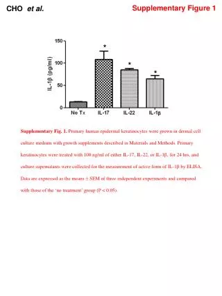
This study examines the impact of cytokine treatments, specifically IL-17, IL-22, and IL-1β, on primary human epidermal keratinocytes. Keratinocytes were cultured and treated with 100 ng/ml of each cytokine for 24 hours, followed by the measurement of active IL-1β levels via ELISA. Additionally, the proportions of neutrophils and dendritic cells in wild-type and caspase-1 knockout mice were assessed, highlighting differences in total cell counts. Results are statistically significant (P < 0.05) across three independent experiments.

Cytokine Treatment Effects on Human Keratinocytes and Immune Cell Proportions in Mice
E N D
Presentation Transcript
Supplementary Figure 1 CHO etal. * * * Supplementary Fig. 1. Primary human epidermal keratinocytes were grown in dermal cell culturemedium with growth supplements described in Materials and Methods. Primary keratinocytes were treated with 100 ng/ml of either IL-17, IL-22, or IL-1, for 24 hrs, and culture supernatants were collected for the measurement of active form of IL-1 by ELISA. Data are expressed as the means SEM of three independent experiments and compared with those of the ‘no treatment’ group (P< 0.05).
Supplementary Figure 2 CHO etal. * * * A B No Tx IL-17 IL-22 IL-1β 47 31 36 6.7 C56BL/6 1.8 5.3 4.2 23 Caspase-1 KO CD11b Gr-1 C No Tx IL-22 IL-1β IL-17 3.8 21 9 15 C56BL/6 2.8 4 3.2 7 Caspase-1 KO CD11c Class II MHC
Supplementary Figure 1 CHO etal. Supplementary Fig. 2. The total cell numbers and the proportion of neutrophils (Gr-1+ , CD11b+ cells) and dendritic cells (CD11c+, class II MHC+ cells) in WT and caspase-1 KO mice. (A) Total cells isolated from mice ears were collected and numbered. Data are expressed as means ± SEM of three independent experiments (* P < 0.05). (B) Cells isolated from ears of mice were stained for 20 min at room temperature with anti-mouse Ly-6G/Ly-6C (Gr-1) (108405, BioLegend), anti-CD11b (101207, BioLegend) to detect neutrophils. (C) Cells were stained with anti-mouse CD11c (117307, BioLegend, San Diego, CA, USA), and anti-mouse I-A/I-E (107605, BioLegend) to detect dendritic cells. Data were acquired on a FACSCalibur system (BD Bioscience) and analyzed using CellQuest software (BD Bioscience).