Rheumatoid Hand Deformities: Pathology Stages and Specific Problems
961 likes | 988 Vues
Understanding the progression of Rheumatoid Arthritis in the hand, from synovitis to joint destruction and deformity. Learn about stages, tendon ruptures, and key issues affecting wrist and DRUJ. Detailed overview with focus on treatment considerations.

Rheumatoid Hand Deformities: Pathology Stages and Specific Problems
E N D
Presentation Transcript
Dr AbhishekShetty RHEUMATIOD ARTHRITIS
PATHOLOGY Stage 1 - Synovitis • Vascular congestion • Proliferation of synoviocytes • Infiltration of subsynovial layers by polymorphs, lymphocytes & plasma cells • Thickening of capsular structures, • Villous formation of synovium, • Cell-rich effusion into joints & tendon sheaths
Stage 2 - Destruction • Persistent synovitis causes joint & tendon destruction • Articular cartilage is eroded 1. Proteolytic enzymes 2. Vascular tissue in fold of synovial reflections 3. Direct invasion of cartilage by pannus of granulation tissue • Joint margin erosion – Granulation tissue – Osteoclastic resorption – Synovial hyperplasia • Tendon sheaths – Similar changes – Tenosynovitis – Invasion of collagen bundles – Partial / complete rupture of tendons
Stage 3 - Deformity • Articular destruction, Capsular stretching, Tendon rupture leads to…. • Progressive instability & deformity of joint • Disruption of normal architecture of hand & wrist • Loss of delicate balance of flexor & extensor forces across adjacent joints of hand-wrist unit
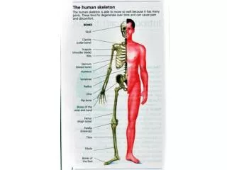
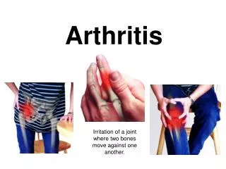
Rheumatoid hand deformities usually are bilateral and symmetrical. Each deformity must be analyzed in detail before surgery is considered. Although combinations of deformities occur, involvement of the fingers, thumb, and wrist is typical. The metacarpophalangeal joints and the wrist are affected early in rheumatoid arthritis, whereas the distal two joints usually are affected later. The metacarpophalangeal is the most important joint affecting finger function in rheumatoid disease. Ulnar deviation with metacarpophalangeal palmar subluxation or dislocation of the finger typifies the rheumatoid hand deformity
RHEUMATOID HAND DEFORMITIES Wrist • Abduction & volar subluxation (radial tilt) • Radial deviation of hand DRUJ • Dorsal Subluxation DRUJ (piano key sign) • Caput Ulnae Fingers / thumb • Ulnar drift deformity (ulnar deviations of digits) • Swan neck & Boutonniere deformity& trigger finger Tendons • Tenosynovitis & Tendon ruptures)
RHEUMATOID HAND DEFORMITIES TENDON RUPTURES JOINT DESTRUCTION COMRESSIVE NEUROPATHIES VASCULITIS
THE WRIST & DRUJ Disturbance of WRIST is common in RA – Initial presentation of wrist unusual ~ 2% – Hand affected 5 x more frequently – Chronic: 90% have functional difficulties due to problems at the wrist KEYSTONE OF THE HAND Pain-free, stable, mobile wrist is necessary for normal function of hand Power generated in finger flexors / extensors – Crosses wrist in order to act fully – Appropriate grasp & motion
THE WRIST & DRUJ Painful, unstable, deformed wrist Impair hand function regardless of status of fingers Wrist deformity Contributes to development of finger deformities Worsens preexistent deformity Compromises surgical correction Unless wrist deformity is preserved/restored Maintaining correction of finger deformity is difficult / impossible
THE WRIST & DRUJ Rheumatoid synovitis of wrist follows predictable patterns Frequently earliest to be involved • Ulnar styloid • Ulnar head • Midportion of scaphoid Progressive synovial proliferation in these areas leads to various patterns of wrist deformity
RHEUMATOID HAND DEFORMITIES Specific Problems at the Wrist 1. Carpal Collapse 2. Translation / translocation of Carpus 3. Volar subluxation 4. Dislocation & supination of carpus 5. DRUJ instability 6. Flexor & Extensor tendon ruptures
COLLAPSE DEFORMITY RADIAL SIDE OF WRIST – Attenuation of Ligaments • Deep radioscapholunate lig. • Radioscaphocapitate lig. – Instability of scaphoid proximal pole – Rotary subluxation of scaphoid – Scaphoid assumes volar flexed position • Secondary loss of carpal height • Radial rotation of carpus &metacarpals on the radius Scapholunate dissociation
ULNAR SIDE OF WRIST – Attenuation of Ligaments • Ulnolunate • Ulnotriquetral – Triangular fibrocartilage – Triquetro-lunate dissociation
TRANSLOCATION & TRANSLATION TRANSLOCATION OF CARPUS • Movement of carpus wholly towards the ulnar side • Rupture of volar radio-luno-triquetral ligaments • Exacerbated by removal of ulnar head TRANSLATION • Radial deviation of carpus • Wrist is in a position whereby fingers are mal-aligned • Leading to ulnar deviation of fingers
VOLAR SUBLUXATION Lunate subluxes from its fossa anteriorly Starts to encroach into carpal tunnel Radius responds by developing shelf of bone Secondary degenerative changes Distortion of carpus & relationship to radius ↑ risk of carpal tunnelsyndrome
SUPINATION OF CARPUS Failure of lunate to remain within its fossa – Rupture of dorsal ulnar carpal ligaments – Dropping of ulnar side of hand – Apparent very prominent ulnar head Which may actually be in its normal anatomic position – Significant distortion of extensor retinaculum – This presses extensor tendons firmly up against the ulnar head • Erosions within ulnar head • Instability of DRUJ • Progressive tendon rupture from ulnar side – Ext Dig Minimus – Ext Dig to 5th, 4th, 3rd , 2nd
SUPINATION OF CARPUS XRAY – Scalloping around ulnar head &distal radius at sigmoid notch – Highly indicative of very unstable DRUJ – Heightened risk of developmentof ruptures of extensor tendons
DISTAL RADIO-ULNAR INSTABILITY Erosion around insertion oftriangular cartilage Attenuation of volar & dorsalulnar carpal ligaments Chronic synovitis with stretching of capsule Attenuation of extensor retinaculum – True dorsal displacement of ulna – Becomes painful (persisting synovitis & crepitus) – Caput ulnae syndrome
CAPUT ULNA SYNDROME • Significant disability • Complain of – Weakness – Pain – Aggravated by forearm rotation
CAPUT ULNA SYNDROME Examination – Prominence of distal ulna – Instability of DRUJ – Limited wrist dorsiflexion – Supination of carpus on forearm – Piano Key Sign • Wrist collapse – Imbalance of extensor tendons – Radial shift of metacarpals – Ulnar deviation of fingers – Important factor in initiating ulnar deviation of MP joints – Recurrence of ulnar deviation following MP joint arthroplasty
PATTERNS OF JOINT DESTRUCTION Radiographic appearance of significant rheumatoid disease Type 1 Ankylotic Type 2 Arthrotic Type 3 Unstable
LARSEN RADIOGRAPHIC SCALE Grading joint involvement in RA Grade O No changes Grade 1 Slight changes,Periarticular swelling Grade 2 Erosions,with definitive joint-space narrowing Grade 3 Medium destructive changes, With erosions & joint spaces poorly defined Grade 4 Severe destructive changes, collapse with significant erosions Grade 5 Mutilating changes
SURGICAL INTERVENTIONS AIMS OF SURGERY To remove diseased synovium via synovectomy To prevent or repair tendon ruptures To provide painless stable wrist joint in functional position. METHODS Synovectomy Distal ulna resection Dorsal stabilization Wrist Arthrodesis Arthroplasty
SYNOVECTOMY To remove diseased synovium before tendon damage occurs. Better results in early stage. Combined with other procedures in later stage.
DISTAL ULNA RESECTION Inflamed radio ulnar joint causes pain in pronation and supination. Diseased ulna roughened by erosive effect of inflammatory synovium rodes the extensor tendons above it. Goal of this surgery to remove this noxious force while maintaining stability
DISTAL ULNA RESECTION Indications DRUJ involvement with pain on pronation and supination or extensor tendon rupture. Contra-indication pre-op evidence of ulnar translocation with displacement of lunate from lunate fossa.
DORSAL STABILIZATION Goal is to stabilise progressive volar and ulnar displacement of radio-carpal joint and remove all diseased synovium. Indications painful synovitis with or without bone destruction , painful minor sublaxation with weakness of grip.
WRIST ARTHRODESIS To correct deformity Obtain a painless wrist To allow the wrist to function in optimal position. Indications advanced fixed volar carpal dislocation , wrist instability caused by severe bony deformity. Contra-indications ipsilateral shoulder and elbow involvement.
ARTHROPLASTY To improve alignment . To retain mobility of the wrist. To provide a stable painless joint. Indications an alternative to arthrodesis when wrist extensors are intact with severe joint destruction , especially in complete upper extremity involvement Contra-indications absent wrist extensor power , severe flexion contracture , dislocated wrist and previous infection with any implant arthroplasty.
METACARPOPHALANGEAL JOINT • MCPJ – Keystones of both longitudinal & transverse skeletal arches of the hand – Frequently site of intense synovitis – Ulnar deviation of digits – Volar subluxation of proximal phalanx – Dislocation of MCPJ
METACARPOPHALANGEAL JOINT FACTORS LEADING TO DEFORMITIES 1. Normal Forces 2. Normal anatomy 3. Pathological changes 1. Normal forces a) Gravity b) Power grasp
METACARPOPHALANGEAL JOINT 2. Normal anatomy a) Asymmetrical shape of metacarpal heads (smaller sloping ulnar condyle) b) Unequal collateral ligament length & their differing orientation c) Asymmetry of intrinsic muscles to the small finger (hypothenars are stronger than 3rd volar interosseous)
METACARPOPHALANGEAL JOINT 3. Rheumatoid Changes – bony erosions of the metacarpal head & base of proximal phalanx leading to ↓ joint stability – Attrition & stretching of the collateral ligaments – Stretching of accessory collateral ligaments, allowing ulnar & palmar displacement of palmar plate & flexor tendons at base of finger – Flexor tenosynovitis with resultant stretching of the flexor tendon sheath pulley system -Rupture of extensor digitorum creating unopposed imbalance at the MCPJ – Contracture of intrinsic muscles,with volar subluxation & ulnar deviation of digits
Surgical procedures that are indicated are intrinsic release or transfer for balance, extensor tendon realignment, and metacarpophalangeal joint synovectomy in mild ulnar drift. In severe ulnar drift, often one or more metacarpophalangeal joints have dislocated. Interposition arthroplasty of the metacarpophalangeal joint reliably relieves pain, maintains stability and alignment, and permits acceptable motion
Subluxation of metacarpophalangeal joints of fingers in severe rheumatoid arthritis. B, Subluxations have been treated by resecting metacarpal heads. Because at surgery articular cartilage of joints was eroded, intrinsic release would have been insufficient treatment.
FINGER DEFORMITIES Deformities of the finger can be caused by the normal forces applied to damaged joints by the extrinsic flexors and extensors, tightness of the intrinsic muscles, displacement of the lateral bands of the extensor hood, rupture of the central slip of the hood, or rupture of the long extensor or long flexor tendons.
FINGER DEFORMITIES Intrinsic Plus Deformity The intrinsic plus deformity is caused by tightness and contracture of the intrinsic muscles. In hands with intrinsic plus deformity, the proximal interphalangeal joint cannot be flexed while the metacarpophalangeal joint is fully extended.
FINGER DEFORMITIES INTRINSIC PLUS DEFORMITY When indicated, intrinsic tightness may be released in conjunction with synovectomy by mobilization of the lateral band.
FINGER DEFORMITIES SWAN-NECK DEFORMITY Swan-neck deformity is described as a flexion posture of the distal interphalangeal joint and hyperextension posture of the proximal interphalangeal joint It is caused by muscle imbalance and may be passively correctable, depending on the fixation of the original and secondary deformities Although usually associated with rheumatoid arthritis, swan-neck deformity may occur in patients with lax joints and in patients with conditions such as Ehlers-Danlos syndrome
Rheumatoid swan-neck deformities of varying severity in all fingers. Metacarpophalangeal subluxation and flexion contractures also are present.
SWAN-NECK DEFORMITY. Terminal tendon rupture may be associated with synovitis of distal interphalangeal joint, leading to distal interphalangeal joint flexion and subsequent proximal interphalangeal joint hyperextension. Rupture of flexor digitorum superficialis tendon can be caused by infiltrative synovitis, which can lead to decreased volar support of proximal interphalangeal joint and subsequent hyperextension deformity. Lateral-band subluxation dorsal to axis of rotation of proximal interphalangeal joint preventing the normal flexion of PIP joint.
Nalebuff, Feldon, and Millender swan-neck deformities Type I deformities are flexible and require flexor tenodesis of the proximal interphalangeal joint, fusion of the distal interphalangeal joint, and reconstruction of the retinacular ligament. Type II deformities are caused by intrinsic muscle tightness and require intrinsic release in addition to one or more of the aforementioned procedures. Type III deformities are stiff and do not allow satisfactory flexion, but do not have significant joint destruction radiographically. These deformities require joint manipulation, mobilization of the lateral bands, and dorsal skin release. Type IV deformities have radiographic evidence of destruction of the joint surface and stiff proximal interphalangeal joints, which usually can be best treated with arthrodesis of the proximal interphalangeal joint
FINGER DEFORMITIES BOUTONNIERE DEFORMITY Caused by underlying joint synovitis which produces an attenuation and lengthening of central slip of extensor digitorum communis tendon. Lateral bands become lengthened as apart of extensor mechanism of PIP joint and are displaced volarwards. Attempted extension at PIP leads to flexion. With increased power n overpull of extensor slip at DIP occurs leading to hyperetension.