Download
1 / 13
130 likes | 202 Vues
Learn how SDS-PAGE separates proteins by mass and how Western blotting detects specific proteins using antibodies. Explore techniques and applications in biochemical research.

E N D
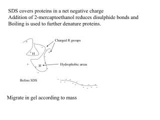
SDS covers proteins in a net negative charge Addition of 2-mercaptoethanol reduces disulphide bonds and Boiling is used to further denature proteins. Charged R groups + - - - + + H - + + Hydrophobic areas H + - - - - Before SDS - - - - - - - - - - - - - Migrate in gel according to mass
Proteins are separated in a ‘discontinuous’ system. Stacking gel has looser pores to allow proteins to line up first. How does an SDS-PAGE gel really work? http://mullinslab.ucsf.edu/Protocols%20HTML/SDS_PAGE_protocol.htm
SDS PAGE gel separates proteins present in a sample All proteins are covered with negatively charged SDS and migrate according to mass Native PAGE gels run under non-denaturing conditions- SDS and 2-mercaptoethanol are omitted from the gel and sample Proteins separate according to charge, size, shape
IgM serum serum Ig What does a Western blot tell you that a protein gel does not? mAb detects light chain Silver stain Western blot Bromage, E. Comp Biochem Physiol B Biochem Mol Biol. 2006 Jan;143(1):61-9. Epub 2005 Dec 1.
Protein blotting • Two major factors affect the efficiency • The elution from the gel -use the lowest percentage of acrylamide that will allow resolution -high molecular weight proteins blot poorly • Efficiency of binding to the membrane • nitrocellulose (not covalently bound) • Polyvinylidene fluoride (PVDF) • Activated nylon
Detection Primary antibody followed by: Radioactive-labelled 125I staphlococcal protein A or streptococcal protein G Enzyme-linked secondary antibodies -horseradish peroxidase (HRP) -alkaline phosphatase-BCIP/NBT BCIP (5-Bromo-4-Chloro-3'-Indolyphosphate p-Toluidine Salt) and NBT (Nitro-Blue Tetrazolium Chloride). Chemiluminescent detection- HRP catalyzes the oxidation of luminol in hydrogen peroxide. Luminol decays by light emission. AP catalyzes the dephosphyorylation of adamantyl-1-2-dioxetane phosphate, resulting in emission of light.
Far western technique Detection of protein-protein interactions using a labelled bait protein
Southwestern blot Figure 7 Distribution of the 52 kDa protein in various mouse tissues as analysed by Southwestern blot analysis Biochemical Journal (1998) 329, 623-629 - www.biochemj.org