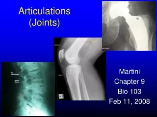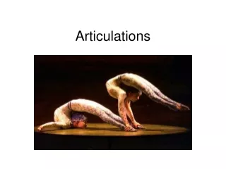
Articulations
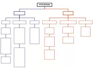
Articulations. “Joints”. Joints. Provide growth and movements to the body. There are two ways that we can classify joints: Functional & Structural. Functional Classification. Synarthroses : immovable joints Ex: sutures of the skull Amphiarthroses : slightly movable

Articulations
E N D
Presentation Transcript
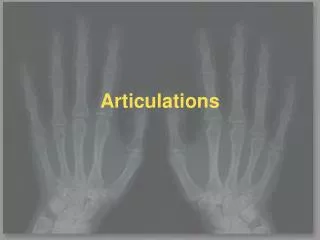
Articulations “Joints”
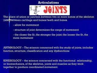
Joints • Provide growth and movements to the body. • There are two ways that we can classify joints: Functional & Structural
Functional Classification • Synarthroses: immovable joints Ex: sutures of the skull • Amphiarthroses: slightly movable Ex: pubic symphysis or intervertebral joints • Diarthroses: freely movable
Flexibility Varies in Diarthrotic Joints • Non-axial: allows only slipping. Plane Joints Ex: wrist • Uniaxial: move in one plane. Ex: elbow • Biaxial: move in 2 planes Ex: saddle joint (thumb) • Multiaxial: move in all planes Ex: ball & socket (shoulder)
Distinguishing Features of Diarthrotic Joints • 1. Articular Cartilage: Hyaline cartilage covers the ends of the bones forming the joint. • 2. Fibrous articular capsule: joint surfaces are enclosed by a capsule of fibrous connective tissue, and the capsule is lined with a smooth synovial membrane =Synovial Joints
3. Joint Cavity: the articular capsule encloses a cavity, which contains synovial fluid. • 4. Reinforcing Ligaments: Capsule is usually reinforced by ligaments.
Now… For the Structural Classifications • But first…… Some Key terms: Ligament: strong, tensile, connective tissue cords that serve the function of uniting bones. Bursae: closed fibrous sacs that are filled with viscous fluid. Frequently located near joints where tendons are subject to frictional forces. Synovial Tendon Sheaths: bursal sacs that surround certain tendons.
3 Basic Structural Types • Fibrous • Cartilaginous • Synovial • Fibrous and Cartilaginous joints lack a joint cavity. • Synovial Joints have a joint cavity.
Fibrous Joints • Permit little or no movement • Synarthroses or Amphiarthroses • Participating bones bound together by fibrous connective tissue. • 3 Specific Types Sutures, Gomphoses, Syndesmoses
Sutures • Bones of the skull • Sutural ligament: thin , intervening layer of fibrous connective tissue. • With age this ligament is transformed into bone.
Gomphoses • Gomphosis: peg and socket type joint. • Synarthrosis • Teeth fixed to maxilla and mandible by gomphoses.
Syndesmoses • Amphiarthroses (slightly movable) • Similar to suture • Ex: distal tibiofibular joint • Common sprain location
Cartilaginous Joints • Lacks a joint cavity • Participating bones held together by intervening cartilage. • Permit little or no movement • 2 Types: Symphyses Synchondroses
Symphysis • Ex: Pubic Symphysis 2 bones held together by interpubic disc consisting of fibrocartilage. Amphiarthrotic. Why? • Ex: Vertebrae seperated by intervertebral disks.
Synchondroses • Temporary joint of hyaline cartilage • Involved in growth • Ex: Epiphyseal cartilaginous plate
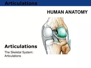
Synovial Joints • Articulating bones are separated by a fluid-containing joint cavity.
Structure of Synovial Joints • Articular cartilage covers ends of bones. • Participating bones bound together by fibrous capsule. It imparts strength and flexibility. Attached to periosteum of participating bones • Synovial Cavity surrounds portions of bones in the formation. • Synovial Membrane: thin membrane that lines all nonarticular aspects of the joint.
Synovial membranes produce synovial fluid. Nutritive and friction reducing properties. • Synovial membrane is not present on articular cartilages. • In certain synovial joints, accumulations of adipose tissue occur within the synovial membrane = articular fat pads = increase surface area of synovial membrane and facilitate distribution of synovial fluid.
Synovial Fluid • Viscous fluid with clear or pale yellow tint. • Contains various cell types: macrophages free synovial cells various white blood cells A cells: possess phagocytic activity. Remove debris and infectious organisms. Produce Hyaluronic Acid (viscosity)
Limit abnormal, excessive motion. Ex: hyperextension of the knee Named according to their location with respect to their attachment. Accessory ligaments: distinct structures that can be located in an intracapsular (ACL, PCL) as well as extracapsular position. Reinforcing Ligaments
Meniscus or Articular Disk • Menisci consist predominantly of fibrocartilage. • Found where articular surfaces lack congruity. • Add to the stability of the joint. • May divide joint cavity into 2 separate compartments, each with own synovial membrane. • Exact function unknown. Proposed: spread of synovial fluid, shock absorption
Types of Synovial Joints • Plane Joints • Hinge Joints • Pivot Joints • Condyloid or Ellipsoidal Joints • Saddle Joints • Ball and Socket Joints
Plane Joint • Participating bones possess flat surfaces of articulation. • Biaxial movement: gliding motions in a single plane but at right angles to each other
Hinge Joint • Uniaxial movement • Collateral ligaments usually provide additional stability. • Ex: Humeroulnar aspect of elbow joint
Pivot Joints (trochoid) • Uniaxial • Permit rotation about a single axis. • Ex: Radioulnar articulation of the elbow.
Condyloid or Ellipsoidal Joints • Condyloid: temporomandibular • Ellipsoidal: biaxial. Radiocarpal joint
Saddle Joints • Biaxial • Variation of ellipsoid type but permits greater flexibility • Ex: carpometacarpal joint of the thumb
Ball and Socket Joint • Spherical head of bone articulates into a cup like surface of another. • Multiaxial movement
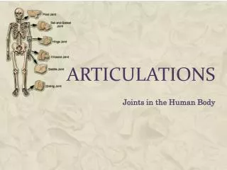
Examples of Joints • A). Knee Joint • B). Hip Joint • C). Shoulder Joint • D). Elbow Joint
Major forms of joint disorders • Dislocation: displacement of a bone from it’s joint. • May be accompanied by injury to ligaments, tendons, and fibrous capsule. • Inflamation: inflammatory involvement of joints is known as arthritis. • Most commonly found in hip, knee, elbow, ankle, wrist, and shoulder.
Gouty Arthritis • Gout is a systemic disorder of uric acid metabolism. • Chronic form of gout is characterized by deposition of uric acid crystals on articular surfaces of joints and by the formation of tophi in fibrous capsule and other connective tissue structures near joint. • Tophi: Uric acid crystals surrounded by cells that are typical of an inflammatory response.
Treatment of Gout • Gout is treatable, and there are ways to keep it from coming back. Treatment usually consists of: • Nonsteroidal anti-inflammatory drugs to relieve symptoms. • Steroidal anti-inflammatory drugs, such as prednisone • While there is no way to guarantee that another attack of gout won't occur, a number of steps can reduce the risk of a future attack. These include: • Taking drugs to slow the production and speed elimination of uric acid • Maintaining a healthy weight, which reduces the pressure on weight-bearing joints and may decrease uric acid levels. Weight loss should be done in a slow, steady way because fasting or rapid weight loss can temporarily raise uric acid levels. • Avoiding too much animal protein. These foods contain purines. • Limiting or avoiding alcohol • Drinking plenty of liquids. Fluids help dilute uric acid in the blood and urine.
Osteoarthritis • Most common form of arthritis. • Articular cartilage deteriorates and becomes eroded. • New bone in the form of “spurs” forms at the periphery. • Some of these may break off and lie free in the synovial cavity. “Joint Mice” • No known cure.