Understanding Brain Development and Cranial Nerves
Dive into the development of the brain and spinal cord, explore different parts like the prosencephalon, mesencephalon, and rhombencephalon, understand the significance of meninges, cerebrospinal fluid, and the different parts of the brain.

Understanding Brain Development and Cranial Nerves
E N D
Presentation Transcript
BRAIN & CRANIAL NERVES The brain and spinal cord develop from ectoderm
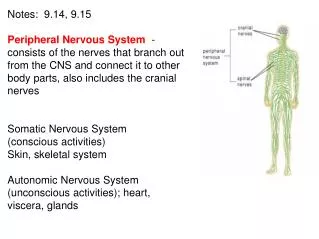
The brain and spinal cord develop from the neural tube • Prosencephalon (Forebrain) • Mesencephalon ( Midbrain) • Rhombencephalon (Hindbrain) • Primary brain vesicles are present around the 3rd week of development
Prosencephalon (forebrain) • Differentiates in 5th week into: • Telencephalon • cerebral hemispheres • Diencephalon • thalamus; hypothalamus • pineal gland
Midbrain • Mesencephalon remains the same • Brainstem = midbrain
Rhombencephalon • Metencephalon • cerebellum, pons • Myelencephalon • medulla oblongata
Meninges • Dura mater • dural septa (extensions): falx cerebri, falx cerebelli, tentorium cerebelli • Arachnoid • Pia mater • Cavities • subarachnoid space, subdural space
4 Ventricles filled with CSF • Lateral ventricles (paired) • Interventricular foramen – connects to 3rd ventricle • Third ventricle • Cerebral aqueduct – connects 3rd and 4th ventricles • Fourth ventricle • Connects with central canal of spinal cord
Cerebrospinal Fluid • Clear, colorless fluid that protects brain • Formed by the choroid plexus • Absorbed by arachnoid villi
Medulla oblongata • Major reflex center for the cardiovascular and respiratory system • vasomotor, vasoconstriction • Pyramids - decussation (crossing) center for motor `tracts • Non-vital center for coughing, hiccuping, swallowing, vomiting • Ascending/descending fibers pass through • Cranial nerves VIII-XII arise here
PONS • Acts as a bridge connecting the spinal cord to the brain • Major relay center for voluntary skeletal movements from the cerebral cortex to the cerebellum • Coordinates with the medulla to regulate respiration • Cranial nerves V-VIII emerge here
Midbrain • Corpora quadrigemina • Visual and auditory reflex centers • Cerebral peduncles - containing large fiber tracts going to and from the brain • Houses the cerebral aqueduct • Cranial Nerves III-IV emerge here
Thalamus • Two halves connected by the intermediate mass • Relay center for ALL sensory cranial and spinal nerves • Interpretation center for crude awareness of pain, temperature and pressure • Location of 3rd ventricle
Hypothalamus • Links the nervous system and endocrine system • Major regulator of homeostasis • Regulates many ANS functions • Regulates appetite, water balance, thirst, body temperature • Emotional part of brain - pleasure, fear, rage
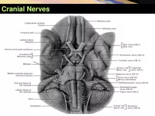
Cranial Nerves • 12 Pairs • 1 Olfactory - smell • 2. Optic - sight • 3. Oculomotor - eye movements • 4. Trochlear - eye movements • 5. Trigeminal - eyes & face, chewing • 6. Abducens - eyes
Cranial Nerves (continued) • 7. Facial - facial expression, taste • 8. Vestibulocochlear - equilibrium, hearing • 9. Glossopharyngeal - tonge & swallowing • 10. Vagus - heart, visceral organs • 11. Accessory - neck & back • 12. Hypoglossal - tongue
Cerebellum • Second largest area of the brain • 2 cerebellar hemispheres • Arbor vitae - branchlike pattern • Vermis - wormlike structure that connects left & right side • Major coordination of skeletal muscle contraction • Assists with posture and balance
Cerebrum • Cerebral cortex - outer layer of gray matter • Two hemispheres separated by longitudinal fissure • Gyri - ridges on surface • Sulci - grooves on surface • Fissures • Septum pellucidum - thin wall between ventricles
Lobes of the Brain • Frontal • Parietal • Occipital • Temporal • Central sulcus • Lateral sulcus
Cerebral Dominance • Left hemisphere • Language • Logic • Math • Right Hemisphere • Artistic • Musical • Creative
Cerebral Cortex Specialization • Motor Areas • Control opposite side of body • Control voluntary motor functions • Sensory Areas • Detect sensations from opposite side of body • Association Areas • Integrate diverse information into purposeful action
Association Areas • Prefrontal Cortex – intellect, learning, & personality • Language Areas • Wernicke’s area – sounding out new words • Brocas’s area – speech • General Interpretation • Visceral Interpretation
Basal Ganglia • Cerebral nuclei • Islands of gray matter located deep within the white matter • Function: controls large automatic skeletal muscle movements and produce dopamine
White Matter • Commissural fibers (corpus callosum) - connect corresponding parts of two hemispheres • Association fibers - connect within the same hemisphere • Projection fibers (higher centers to lower ones)
Limbic System • Includes part of thalamus, hypothalamus, and cerebrum • Emotional brain • Associated with memory • Involuntary behavior for survival • Pleasure and pain centers - fear, sorrow, affection
Reticular Formation • RAS System - reticular activating system • Network of gray matter extending from the medulla, pons, midbrain into the cortex • Maintains consciousness • Awaken from sleep • Alerts brain of incoming sensory signals