Download
1 / 57
570 likes | 762 Vues
Pediatric OMT Swimmers Module. American College of Osteopathic Pediatricians Robert Hostoffer, DO,FACOP, FAAP. edited by Eric Hegybeli, DO, FACOP. questionnaires by Michael Rowane, DO. Background: Andrew Taylor Still, MD, DO. Born in Virginia in 1828

E N D
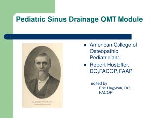
Pediatric OMT Swimmers Module • American College of Osteopathic Pediatricians • Robert Hostoffer, DO,FACOP, FAAP • edited by • Eric Hegybeli, DO, FACOP questionnaires by Michael Rowane, DO
Background: Andrew Taylor Still, MD, DO • Born in Virginia in 1828 • The son of a Methodist minister and physician. • At an early age, Still decided to follow in his father's footsteps as a physician. • After studying medicine and serving an apprenticeship under his father, Still became a licensed M.D. in the state of Missouri. • Completed additional coursework at the College of Physicians and Surgeons in Kansas City, Missouri [Early 1860's] • Union Army Surgeon during the Civil War.
Background: Andrew Taylor Still, MD, DO • After the Civil War and following the death of three of his children from spinal meningitis in 1864, Still concluded that the orthodox medical practices of his day were frequently ineffective, and sometimes harmful. • He devoted the next ten years of his life to studying the human body and finding better ways to treat disease. • Discovered Osteopathy in 1874
Background: Andrew Taylor Still, MD, DO • His research and clinical observations led him to believe: • The musculoskeletal system played a vital role in health and disease • The body contained all of the elements needed to maintain health, if properly stimulated. • By correcting problems in the body's structure, through osteopathic manipulative treatment, the body's ability to function and to heal itself could be greatly improved.
Background: Andrew Taylor Still, MD, DO • Promoted the idea of preventive medicine • Endorsed the philosophy that physicians should focus on treating the whole patient, rather than just the disease. • http://www.aacom.org/OM/history.html
Background An average competitive swimmer can complete between 6,000 yrads in a two hour session. Approximately 20-40 miles per week. Approximately 1 million stroke cycles per year.
Risk Factors Laxity vs. Instability Poor stroke mechanics Excessive fatigue Improper or excessive stretching Improper weight training Excessive use of kick boards
Laxity vs. Instability Laxity: normal; pain free ROM of a joint Instability: Pathologic subluxation or dislocation resulting in pain or functional impairment.
Many swimmers have joint laxity Laxity may foster glenohumoral instabilty leading to impingement
Stroke Mechanics Excessive stress Fatique Increased Drag Poor Technique
Stroke Analysis • Wringing out of supraspinatus • Crossing Midline • Flat Body Positioning • Promote impingement • Hawkin’s Position • Dropped elbow • First sign of fatigue or pain • Minimizes time in impingement position
Modifying Stoke Mechanics • Breathe bilaterally • Increases body roll • Maitain high elbow during recovery • Lessens demand on scapular stabilization musculature
Stretching • Swimming promotes strengthening of the glenohumoral internal rotators and pectoral musculatures • Most swimmers excessively stretch the anterior capsule • Buddy stretches
Who should stretch? • Swimmers without pain
Who should not stretch? Swimmers with multidirectional instability or shoulder pain A gentle warm up should replace pre-workout stretching
Weight Training • Minimize abduction and external rotation • Lat pulldowns, military press, shoulder abduction • Emphasize scapular retractors, glenohumoral external rotators and core musculatures
Kicking Excessive use of board promotes impingment Neer’s position
Neer’s Stages Stage I refers to those with edema and hemorrhage; Stage II refers to those with fibrosis and tendinitis; Stage III refers to those with tear of the rotator cuff, ruptured biceps or bone excrescence
Treatment • 3 phases • Acute • Recovery • Functional retraining
Acute phase • RICE • NSAIDs • Possible subacromial injections • Avoiding impingment positions • OMT A/AAROM • Modalities • Ultrasound • TENS
Recovery • Restore normal AROM-OMT • Restore strength and endurance to shoulder stabilizing musculature and rotator cuff • High repetition of low weight or low resistance elastic bands
Functional Phase • Entrance Criteria • Normal Shoulder AROM • Rotator cuff strength at least 4/5 • Normal and functional kinetic chain • Goals • Sports specific training • No muscle in isolation
Swim Bench • Isokinetic exercise • Return to pool with gradual increase in yardage/intensity • Plyometric exercises • Neuromuscular intergration • Very high force generation • Complementary muscle group coordination and strengthening
Surgery • Indications • Failure of conservative management • Paresthesias, dead arm • Significant instability • Difficulty with ADLs ( activity of daily living) secondary to pain
Surgical techniques Inferior Capsular Shift Anterior capsulolabral reconstruction Arthroscopic repair Thermal Capsular Shrinkage
Swimmer Injury Painful arc/rotator cuff pain in the shoulder of a swimmer can occur in any of the following movements: 1. Adduction of the arm at the shoulder 2. When this movement is blocked 3. Flexion of the arm at the shoulder 4. When this movement to left or right is blocked.
The acromioclavicular joint may develop degenerative arthritic changes, particularly from damage in resistance weight training.
Arthritis of the glenohumeral joint may be seen in the masters age group, though it is rare in the young.
breast-stroke knee the alignment of the knee centre relative to the hip centre during the start of the breast-stroke kick affects the development in the medial collateral ligament and capsule. The optimum initiating position from the breast-stroke kick is with the hip and knee centres aligned. When the knee centre is narrow or wide of the hip centre, it causes increased stress on the medial collateral joint structures. Exceeding the elastic limits of the ligament will cause damage and injury. In young swimmers, this form of stress could open growth plates of the femur and tibia and cause micro-injury which will result in inflammation and thus seriously impair training.
Patella Injury There is a high risk of the patella riding laterally during the breast-stroke kick. This is magnified when the patella tendon attachment site at the tibial tubercle is placed in an extremely rotated position. Weakness of the vastus medialis can decrease effectiveness in ensuring central tracking of the patella. If dislocation occurs, surgery is almost certain.
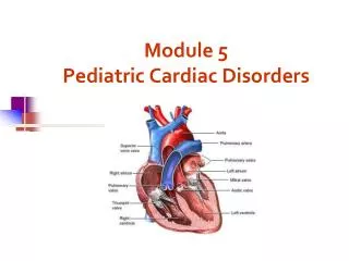
Shoulder Anatomy • Acromiolclavicular joint • Rotator Cuff Muscles: • Supraspinatus • Infrspinatus • Teres Minor • Subscapularis • Biceps muscles • Humerus
Swimmer’s Shoulder Subacromial impingement ( swimmers shoulder ) is a condition that affects the athlete's abililty during the catch phase, early to mid-pull, and through arm adduction in the recovery phase. It is an inflammation of the supraspinatus located on the long head of the biceps tendon. Most often it is a result of incorrect form and overuse.
Swimmer Shoulder Causes Poor Stroke Technique : Improper stroke technique can result in joint and muscle imbalance. Unilateral Breathing : Most swimmers are comfortable breathing in one direction, this results in muscle imbalance in swimmers. Overuse : These are chronic injuries that occur because of repeated stress to the muscles, tendons and joints.
The seven-step Spencer shoulder technique: • Step 1—extension with elbow flexed; • step 2—flexion with elbowextended; • step 3—compression circumduction; • step 4—circumductionwith traction with elbow extended; • step 5a—abductionwith internal rotation with elbow flexed; • Step 5b- adduction and external rotation • step 6—adduction and internal rotation with upper extremity behind the back; • step 7—stretching tissues and pumping fluids with the arm extended
step 6—Abduction and Internal Roatation with the upper extremity behind the back
step 7—stretching tissues and pumpingfluids with the arm extended
Protective Stretches (2 reps – hold for 10-20 seconds each): 1) Put both arms overhead in the streamlined position, then lean first to the left side as far as possible, then to the right. 2) Put both arms behind your back, fingers interlaced, and slowly, steadily raise your arms upward behind you as far as possible. 3) Put one arm across your body so that the shoulder is under your chin and hand, forearm and upper arm are parallel to the ground. Without turning your body, use your other hand to pull the arm close as close to your chest as possible.
Innervation Table Organ/System Parasympathetic Sympathetic Ant. Chapman's Post. Chapman's EENT Cr Nerves (III, VII, IX, X) T1-T4 T1-4, 2nd ICS Suboccipital Heart Vagus (CN X) T1-T4 T1-4 on L, T2-3 T3 sp process Respiratory Vagus (CN X) T2-T7 3rd & 4th ICS T3-5 sp process Esophagus Vagus (CN X) T2-T8 --- --- Foregut Vagus (CN X) T5-T9 (Greater Splanchnic) --- --- Stomach Vagus (CN X) T5-T9 (Greater Splanchnic) 5th-6th ICS on L T6-7 on L Liver Vagus (CN X) T5-T9 (Greater Splanchnic) Rib 5 on R T5-6 Gallbladder Vagus (CN X) T5-T9 (Greater Splanchnic) Rib 6 on R T6 Spleen Vagus (CN X) T5-T9 (Greater Splanchnic) Rib 7 on L T7 Pancreas Vagus (CN X) T5-T9 (Greater Splanchnic), T9-T12 (Lesser Splanchnic) Rib 7 on R T7 Midgut Vagus (CN X) Thoracic Splanchnics (Lesser) --- --- Small Intestine Vagus (CN X) T9-T11 (Lesser Splanchnic) Ribs 9-11 T8-10 Appendix T12 Tip of 12th Rib T11-12 on R Hindgut Pelvic Splanchnics (S2-4) Lumbar (Least) Splanchnics --- --- Ascending Colon Vagus (CN X) T9-T11 (Lesser Splanchnic) R Femur @ hip T10-11 Transverse Colon Vagus (CN X) T9-T11 (Lesser Splanchnic) Near Knees --- Descending Colon Pelvic Splanchnic (S2-4) Least Splanchnic L Femur @ hip T12-L2 Colon & Rectum Pelvic Splanchnics (S2-4) T8-L2 --- ---