Download
1 / 39
400 likes | 547 Vues
The cells that make us who we are How neurons communicate with one another. Communication within the Nervous System Chapter 2. The Cells That Make Us Who We Are. How many are there? Neurons : 100 billion Make up 10% of brain volume Glial cells : Many more! Make up 90% of brain volume

E N D
The cells that make us who we are How neurons communicate with one another Communication within the Nervous SystemChapter 2
The Cells That Make Us Who We Are • How many are there? • Neurons: 100 billion • Make up 10% of brain volume • Glial cells: Many more! • Make up 90% of brain volume • Neurons: • convey sensory information to the brain; • carry out the operations involved in thought and feeling; • transmit commands out to the body to control muscles and organs. ◊
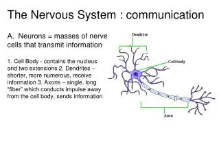
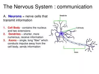
The Cells That Make Us Who We Are • Cell body or soma • Contains the nucleus • The nucleus contains the genetic material (DNA). • Contains organelles in cytoplasm • These convert nutrients to fuel, construct proteins, and remove waste. • Dendrites • Extensions that branch out from the cell body • Receive information from other neurons and sensory cells • Axon • Extends some distance from the cell body • Carries information to other neurons and to organs ◊
The Cells That Make Us Who We Are • Terminals or end bulbs are swellings found at the end of the axon. • Neurotransmitters are chemical substances found inside the terminals. • The neurotransmitters communicate with: • other neurons; • muscles; • organs. ◊
The Cells That Make Us Who We Are • Three major types of neurons that vary by shape: • Multipolarneuron (like the neuron in the previous slide) • Unipolarneuron • Bipolarneuron
The Cells That Make Us Who We Are • Three major types that vary by function • Motorneuron • Motor neurons are multipolar. • Sensoryneuron • Either bipolar or unipolar. • Interneuron • Multipolar • ◊
The Cells That Make Us Who We Are • The most critical factor in the neuron’s ability to communicate is the cell membrane. • Composition of the cell membrane • Lipid molecules and proteins make up the cell membrane. • Lipid molecule heads are attracted to the fluid in and outside the cell; the tails are repelled by liquid. • Some of the lipids orient their heads toward the extracellular fluid and some orient toward the intracellular fluid. • This creates a double-layer membrane. • The cell membrane holds the cell together and controls the environment in and around the cell.
The Cells That Make Us Who We Are • The neuron’s membrane varies in permeability. • Water, oxygen, and carbon dioxide pass freely • Many other substances are barred from entry. • Others can pass through protein channels in the membrane under certain circumstances. • Polarization results from this selective permeability of the membrane. • Polarization is the difference in electrical charge between the inside and outside of the cell. • This difference in electrical charge is referred to as avoltage.
The Cells That Make Us Who We Are • The resting potential is the difference in charge between the inside and outside of the membrane when the neuron is at rest. • Resting potential may be anywhere between -40 and -80 millivolts (mV) in different neurons. • The typical neuron’s resting potential varies around -70 mV. • The resting potential is caused by unequal distribution of ions on either side of the membrane. • The fluid outside the cell contains mostly sodium (Na+) and chloride (Cl-) ions. • The fluid inside the cell contains mostly potassium (K+) ions and organic anions (A-). • There are more negative ions inside the membrane and more positive ions outside the cell.
The Cells That Make Us Who We Are • There are two main forces that determine the location and movement of the ions. • Force of diffusion: ions move from area of high to low concentration. Where would chloride ions move based on diffusion? • Electrostatic pressure: ions are repelled from the side of the membrane with the same charge and attracted to the side with the opposite charge. Where would sodium ions move based on electrostatic pressure? ◊
The Cells That Make Us Who We Are • Why is the membrane polarized? • The organic anions (A-) are too large to leave the cell. • The chloride ions are repelled by the organic anions. • Sodium and potassium channels are closed; only a few of those ions pass through. • Sodium and potassium ions that do leak through are returned by the sodium-potassium pump. • These forces and events combine to maintain the resting potential around -70 mV. ◊
The Cells That Make Us Who We Are • Excitatory signals received by the neuron cause a partial depolarization, or hypopolarization, in a small area of the membrane. • The depolarization is caused by a change in ion balance, which also affects the adjacent membrane. • This spreading depolarization diminishes over distance, so it is often referred to as a local potential. • At the axon, depolarization that reaches threshold (around -60 mV) will cause sodium channels to open, triggering an action potential. ◊
The Cells That Make Us Who We Are • The action potential is an abrupt change in membrane potential that allows the neuron to communicate over larger distances. • Sodium ions enter the open sodium channels due to force of diffusion and electrostatic pressure. • The rate of entry of sodium is 500 times greater than normal. • The result is complete depolarization. • Polarity “overshoots” to around +30 to +40 mV, making the interior of the cell temporarily positive. • Sodium channels close at the peak of the action potential and no further depolarization is possible. ◊
The Cells That Make Us Who We Are • Near the peak of the action potential, potassium channels open. • Potassium ions move out due to diffusion. • Potassium also moves out due to electrostatic pressure because the inside of the cell is temporarily positive. • The membrane returns to near resting potential. • This entire event takes approximately 1 millisecond. • Because nearby sodium channels open, a new action potential is triggered at the adjacent patch of membrane. ◊
The Cells That Make Us Who We Are • Movement of action potentials down the axon is not a flow of ions but a chain of events. • When the action potential reaches the terminals it passes the message on to the next cell “in line”. • The local potential is a graded potential, but the action potential follows the all-or-none law. • All-or-none means that the action potential always occurs at full strength and does not vary with stimulus intensity. • The action potential also is nondecremental; it does not decrease over distance. • This makes it possible for the message to travel over long distances.
The Cells That Make Us Who We Are • Absolute Refractory Period • When the sodium channels close during the action potential, that part of the axon cannot fire again. • This limits how frequently the neuron can fire. • This also prevents backward spread of depolarization, so action potentials move only toward the terminals. • Relative Refractory Period • Potassium channels remain open; continued K+ outflow polarizes the membrane beyond the resting potential. • A stronger stimulus is required to trigger an action potential. • Rate law: Stronger stimuli trigger new action potentials earlier in recovery, so the axon encodes intensity as rate of firing.
The Cells That Make Us Who We Are • Neurotoxins affect ion channels involved in the action potential. • The puffer fish produces tetrodotoxin, which blocks sodium channels. • Scorpion venom keeps sodium channels open, prolonging the action potential. • Beneficial drugs affect these ion channels as well. • Local anesthetics block sodium channels. • Some general anesthetics work by opening potassium channels. • Ion channels can be modified to control neurons by light. • This allows greater precision in stimulating neurons and identifying pathways in the brain.
The Cells That Make Us Who We Are • Glial cells produce myelin, a fatty tissue that surrounds axons, providing insulation and support. • CNS: oligodendrocytes make the myelin. • PNS: Schwann cells make the myelin. • Myelin increases the conduction speed in axons. • Myelinated axons have gaps callednodes of Ranvier; action potentials occur only at the nodes. • Transmission between nodes (under the myelin) is by local potential, which moves faster than action potentials and uses less energy. • Because the action potential “jumps” from node to node this is calledsaltatory conduction. • Multiple sclerosis is a disease in which myelin is destroyed, resulting in reduction of conduction speed.
The Cells That Make Us Who We Are • Other Glial Functions • During development, glial cells provide scaffolds for neurons to migrate to their final destinations. • Glial cells also respond to injury and disease by removing cellular debris. • Glial cells provide energy to neurons. • When glial cells are present, neurons make seven times as many connections with other cells • As behavioral complexity increases, the ratio of astrocytes (a type of glial cell) to neurons also increases. ◊
How Neurons Communicate With One Another • A synapse is the connection between the presynaptic neuron and postsynaptic neuron. • The neurons are separated by a space orcleft. • The earlier view was that the nervous system was a continuous web. • Using Golgi staining, which stains only a portion of neurons, Cajal was to see that each neuron is an individual cell. • At that time, physiologists also believed neurons used electric current to communicate across the synaptic cleft. ◊
How Neurons Communicate With One Another • Loewi’s Discovery of Chemical Transmission • Loewi isolated two frog hearts (heart A and heart B). • Stimulating the vagus nerve of heart A slowed the heart. • Salt solution collected from heart A caused heart B to slow. • Stimulating the accelerator nerve caused heart A to beat faster. • Transferring this salt solution caused heart B to beat faster. • Loewi concluded that each nerve released a different chemical into the fluid—one excited the heart and one inhibited it. • This experiment demonstrated synaptic transmission by chemical neurotransmitters (though electrical transmission also occurs).
How Neurons Communicate With One Another • Transmitters are stored in vesicles in the axon terminals. • When the action potential arrives at the terminals, calcium channels open and calcium enters the cell. • This causes vesicles to fuse with the membrane, which results in release of transmitter into the cleft. • Transmitters cross the cleft and influence receptors on the postsynaptic neuron. ◊
How Neurons Communicate With One Another • There are two major types of receptors on the postsynaptic cell. • Ionotropic receptors • cause ion channels to open, which • has a direct and rapid effect on the neuron. • Metabotropic receptors • open channels indirectly, • producing slower but longer-acting effects. • Synaptic transmission takes a couple of milliseconds, so it is slower than axonal transmission. ◊
How Neurons Communicate With One Another • Activation of receptors on the postsynaptic cell has two possible effects on the membrane potential. • Hypopolarizationcreates anexcitatory postsynaptic potential (EPSP). • An EPSP opens sodium channels. • This makes the postsynaptic neuron more likely to fire. • Hyperpolarization creates aninhibitory postsynaptic potential (IPSP). • An IPSP opens potassium or chloride channels or both. • This makes it less likely an action potential will occur. ◊
How Neurons Communicate With One Another • EPSPs and IPSPs are graded potentials. • Their effects can accumulate over a short time, an event referred to as temporal summation. • Spatial summation combines inputs arriving at different locations on the dendrites and cell body. • The neuron acts as • an information integrator, by the processes of summation, and • adecision maker, by combining excitatory and inhibitory inputs algebraically, determining whether to fire. ◊
How Neurons Communicate With One Another • Usually, the transmitter’s effects must be terminated to allow frequent responding and to prevent it from affecting nearby synapses. • Reuptake takes the transmitter back into the terminals to be used again. • Glial cells can reabsorb transmitters at some synapses. • Acetylcholine is broken down by acetylcholinesterase. • Some drugs work by enhancing or reducing transmitter effects. For example: • some antidepressants block reuptake of transmitters; • others prevent their degradation.
How Neurons Communicate With One Another • Synaptic activity is regulated in several ways. • One process occurs via axoaxonic synapses. • Presynaptic inhibition decreases the release of transmitter. • Presynaptic excitation increases the release of transmitter. • This regulation occurs by affecting calcium entry into the terminal. • Autoreceptorssense the amount of transmitter in the cleft and cause the presynaptic neuron to reduce excessive output. • Glial cells • prevent transmitter from spreading to other synapses; • absorb and recycle transmitter for the neuron’s reuse; • release glutamate to regulate presynaptic transmitter release.
How Neurons Communicate With One Another • Having a variety of transmitters multiplies the effects that can be produced at the synapse. • Different subtypes of receptors add even more complexity. • For example, nicotine acts at nicotinic and muscarinic receptors for acetylcholine. • Some neurons release more than one transmitter: • a fast-acting transmitter and a slower-acting neuropeptide; • or two fast-acting transmitters; • or an excitatory transmitter and an inhibitory transmitter.
How Neurons Communicate With One Another • Some representative neurotransmitters: • Acetylcholine • Monoamine transmitters • Examples: dopamine, norepinephrine, and serotonin • Amino acid transmitters • Examples: glutamate, GABA, and glycine • Peptide transmitters • Examples: endorphins, substance P, neuropeptide Y • Gaseous transmitters • Examples: nitric oxide, carbon monoxide ◊
How Neurons Communicate With One Another • Neuronal coding and neural networks add further complexity to neural processing. • Trains of neural impulses encode additional information in the intervals between spikes and length of bursts. • Some information is encoded by segregating it to specialized pathways known as “labeled lines.” • In the brain, information is integrated and processed in complex neural networks. • One way of studying these networks is to simulate their activity with computer-basedartificial neural networks. ◊