Comparative Anatomy Integument
460 likes | 1.31k Vues
Comparative Anatomy Integument. Kardong Chapter 6 Part 8. Integument. Epidermis derived from ectoderm Gives rise to glands Dermis derived from mesoderm. Figure 8.1. Figure 8.3. Amphibian skin showing mucous and poison glands. Figure 8.2. Poisonous Dart Frog. Modifications.

Comparative Anatomy Integument
E N D
Presentation Transcript
Comparative AnatomyIntegument Kardong Chapter 6 Part 8
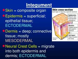
Integument • Epidermis derived from ectoderm • Gives rise to glands • Dermis derived from mesoderm Figure 8.1.
Figure 8.3. Amphibian skin showing mucous and poison glands. Figure 8.2. Poisonous Dart Frog.
Modifications • Presence or absence of bone in dermis • Glands in aquatic forms • Specializations in epidermis of land dwellers Figure 8.4. African hairy frog with specialized hairs acting as auxiliary respirator organs.
Functions of the Skin • Protection • Respiration • Temperature Control • Nourishment of Young • Locomotion and reproductive structures Figure 8.5. African clawed frog (Zenopuslaevis) was used for pregnancy test and spread chytrid fungus around the world.
Fish Skin • No stratum corneum • Many unicellular glands • Like goblet cells, secretes mucus • Photophores in deep-sea fishes Figure 8.6.
Dermal Scales • Dermal bone plates became skull surface bones • Ancient armor • Rhomboid scales • Modern fish • Cycloid and ctenoid scales • Placoid and ganoid scales Figure 8.7. Ostracoderm. Figure 8.8. Cycloid, ctenoid, placoid, and ganoid scales of modern fish.
Dermal Scales (cont.’d) • Ctenoid scales • Growth rings or annuli • No epidermal scales • Scales are dermal Figure 8.10.
Amphibian Skin • Loses dermal scales • Exception: Caecilians and some primitive frogs • Epidermal multicellular glands abundant • Stratum corneum (contains Leydig cells to resist entry of bacteria and viruses) Fig. 8.11. (book fig. 6.12).
Integumentary Gland Type • Simple tubular • Plethodontid mental glands associated with courtship • Simple coiled tubular • Sweat glands • Simple branched tubular • Female plethodontid- spermatheca • Simple alveolar (acinar) • Mucous glands Figure 8.12.
Compound tubular • Mammary glands of monotremes • Compound branched alveolar • Mammary glands of placentals • Courtship glands Figure 8.13.
(a) Simple tubular, (b) Coiled tubular, (c) Simple branched tubular, (d) Compound tubular, (e) Alveolus of simple saccular gland, (f) Simple branched saccular, (g) Compound alveolar Figure 8.14. Morphological varieties of multicellular glands.
Plethodontid Salamanders - Mating • Internal fertilization • Male: mental glands on chin, cloacal glands to form spermatophore, and caudal courtship glands • Female: spermatheca for sperm storage • Glands secrete pheromones (a) Figure 8.15. Salamander spermatophore (book figure 14.40). (b) Figure 8.16. (a) Plethodon (b) spermatophore.
Glands Associated with Mating (a) (b) (c) Figure 8.17. (a) mental glands, (b) cloaca, (c) nasolabial groove.
Modes of Secretion • Merocrine • Holocrine • Apocrine Figure 8.18. Mammalian skin.
Merocrine • Cell body not injured • Release particles by exocytosis • Most sweat glands in mammals • Holocrine • Cell body discharged with contents • Whole cell dies • Sebaceous glands (a) (b) Figure 8.19. (a) merocrine and (b) holocrine glands.
Apocrine • Cellular products gather on surface then pinched off • Apical portion pinched off • Axillary sweat glands Figure 8.20. Apocrine gland.
Reptile Skin • Few glands (dry skin) • Thick stratum corneum with modifications • Epidermal scales Figure 8.21. Desert horned lizard.
Some reptiles have remnants of dermal armor (osteoderms) • Osteoderms beneath some epidermal scales • Gastralia- large osteoderms • Alligator and skinks • True dermal bones • Turtles Figure 8.22. Osteoderms (dermal plates) of alligator.
Turtles • Shell of dermal bone • Carapace (shell) – dorsal • Plastron- ventral • Mesoplastron additional bone on primitive, extinct turtles • Nuchal- diagnostic bone Figure 8.23. Turtle shell.
Reptile Skin • Turtles have epidermal scutes- large epidermal scales • Snakes have scutes on belly • Spikes and spines are epidermal (a) (b) Figure 8.24. Snake belly scutes (a) and white bony plate of turtle with scutes removed.
Reptile Integumentary Glands • Femoral pores • Occur ventrally, waxy excretion • Many lizards and snakes have scent or cloacal glands • Snakes use forked tongue to pick up scent (Jacobson’s organ) Figure 8.25. Prairie Rattlesnake. Figure 8.26. Jacobson’s organ.
Musk Glands • Scent glands • Along carapace in turtles - Rathke’s glands • Under lower jaw in crocodiles • Musk deer • Take secretions to make perfume Fig. 8.27. Turtle Rathke’s glands.
Skin of Birds • Few epidermal scales • Legs and beak • Dermal scales are absent • Claws- diversified • Few glands • Uropygial gland- preening gland • Dermal scales absent Figure 8.28. Feather type (see book figure 6.15).
Feathers • Modification of reptilian scales • 3 types • Contour- flight feather • provides wing shape • Down- beneath contour feather • Filoplume- long shaft • lost its vane Figure 8.29.
Skin of Mammals • Modifications of stratum corneum • Hair, claws, nails, hooves • Hair • Like filoplume feather and lack detail • Vibrissae • Specialized hairs • Tactile in function Figure 8.30. Vibrissae of harbor seal.
Skin of Mammals (cont’d.) Figure 8.31. Cross sections of mammalian skin.
Cornified Structures • Baleen Plate • Toothless whale’s horny sheets of oral ectoderm • Not bone • Used for filter feeding • Tori pads • Epidermal pads Figure 8.32. Products of stratum corneum; tori.
Horns • Horns • In bovid family • Outgrowth of dermal core • Unbranched • Covered by epidermal horny, keratinized sheath • Permanent Figure 8.33. Bovine horn.
Antlers • Antlers and horns of giraffe • Deer • Dermal bones • Dermal bone of antler attaches to skull bone • Shed annually • Outside layer is highly vascularized Figure 8.34. Antler.
Figure 8.35. (a) horns and (b) antlers (see book figure 6.26).
Dermal Pigments • Chromatophores • contain pigment granules • Melanophores (brown) • Melanin granules within melanosomes • Lipophores (yellow and red) • Iridophores or guanophores (iridescent) - Contain reflective guanine crystals