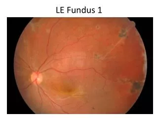
normal fundus
normal fundus. ophthalmoscopy. Indirect. Direct. normal fundus. normal fundus. normal fundus. normal fundus. Optical Coherence Tomography (OCT) uses a light beam the same way that B-scan ultrasonography uses a sound beam to image the retina in a microscopic slice.


normal fundus
E N D
Presentation Transcript
ophthalmoscopy Indirect Direct
normal fundus Optical Coherence Tomography (OCT) uses a light beam the same way that B-scan ultrasonography uses a sound beam to image the retina in a microscopic slice.
Congenital fundus anomalies Opaque retinal nerve fibers- myelinated retinal nerve fibers; bright, white patch adjanced to the disc often obscuring the retinal vessels running in the white patch.
Congenital fundus anomalies • Opaque retinal nerve fibers
Congenital fundus anomalies • Opaque retinal nerve fibers
Congenital fundus anomalies Myopic crescent- 1. white with black pigmented borders rimof atrophic choroid revealing the underlying white sclera in a crescent shape; 2. usually temporal to the disc but may completely surround it; 3. in pathological myopia often associated with myopic choroidoretinal degeneration.
Congenital fundus anomalies • Myopic crescent
Senile changes Age related macular degeneration (AMD) 1. the most common cause of registrable blindness in Western countries; 2. bilateral gradual deterioration of central vision over several years (sometimes sudden), often with symptoms of distortion; 3. the earliest mainifestation, risk factor of AMD - drusen (small yellow spots in macular region, associated with pigment speckling, consist of hyaline, between the retinal pigment epithelium and Bruch`s maembrane, usually cause no visual symptoms).
Senile changes Age related macular degeneration (AMD) 4. non- exudative (dry or atrophic): bilateral progressive atrophy of RPE and the choriocapllares in the macular region secondary to arteriosclerotic degeneration of choroidal vessels; speckled pigmentation followed by the apperance of areas of retinal atrophy with visibility of choroidal vessels; typically slow gradual to modearte loss of vision;
Senile changes Age related macular degeneration (AMD) 5. exudative (dry or atrophic): in early stages retinal oedema (distortion of central vision); less common than non- exudative but causes more severe visual loss!!!; two important features- detachment of RPE and choroidal neovascularisation which may haemorrhage and leads to a fibrous disciform scar at the macula;
Senile changes Wet AMD is associated with new blood vessels (neovascularization) that originate in the choroid and break through Bruch's membrane and the RPE layer. OCT image below indicates the RPE layer that has been broken through by the choroidal new blood vessels. Age related macular degeneration (AMD)
Senile changes Age related macular degeneration (AMD)
Senile changes The end result is scarring and a loss of retinal function in the area affected. Ninety percent of the cases of severe vision loss from AMD results from wet AMD. Age related macular degeneration (AMD)
Senile changes- Amsler grid as it might appear to someone with age-related macular degeneration.
Senile changes Agerelatedmaculardegeneration (AMD) Treatment Non- exudative: no treatment Exudative: argon laser photocoagulation to destroy a choroidalneovascularmembrane, photodynamictherapy- PDT (Visudine), Injectionsintovitreous body (steroids- Triamcinolone, antineovascularizationagents- Macugen, Lucentis, Avastin), nutrientsupplements (vitamin C and E, zinc, cuprum, betacarotene, lutein).
Senile changes Macular Hole generally middle- aged woman; sudden reduction in central vision to around the 6/60 level; small round retinal hole centered on the fovea; Vitreous traction; Treatment- vitrectomy
Senile changes Myopic Degeneration High myopia= large eyeball with all retinal layers stretched and thinned; primary choroidal atrophy may affect the macular region, or breaks in Bruch`s membrane (laquer cracks) through which choroidal neovascularisation and subsequent disciform scarring may develop in a similar manner to AMD
Retinal detachment Separation of the retina from its pigment epithelial layer. The separation occurs at this site for embryological reasons: the two walls of the embryonic optic vesicle become apposed and form respectively the RPE and neuroretina.
Retinal detachment rhegmatogenous RD - most common type of RD; - secondary to a tear or hole in the retina, which often arises as a consequence of posterior vitreous detachment
Retinal detachment non- rhegmatogenous RD (exudative and tractional) - an ucommon type of RD; - there is no defect in retina; - result of exudative processes beneath the retina (e.g. scleritis or choroidal neoplasm) or as result of vitreous tractional forces pulling the retina forward (e.g. proliferative diabetic retinopathy).
Retinal detachment risk factors - age (over 50); - high myopia; - trauma (younger patients); - systemic conective tissue disorders (Marfan`s syndrome)
Retinal detachment symptoms - sudden onset of floating specks or spots associated with flashes of light (typical for posterior vitreous detachment and/ or retinal tear formation); - „shadow” or „curtain” in the visual field that gradually extends to cover the whole visual field (the same day or days or weeks later);
Retinal detachment signs - visual field loss corresponding to the area of detached retina (the temporal retina generally detaches first- nasal field defect); - reduction in visual acuity (if the macular region becomes detached);
Retinal detachment signs - grey retinal folds which quiver as the eye moves; - the blood vessels on the detached retina have deeper red colour than normal; - easy contrast between the normal fundus colour and the greyish detached part of retina;
Retinal detachment signs - retinal break will be seen at the periphery of detached fundus in the form of a tear (arrowed or „U” shaped), hole or dialysis (typical for traumatic RD)