Fundus camera
Fundus camera. A non- mydriatic Topcon retinal camera

Fundus camera
E N D
Presentation Transcript
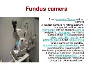
Fundus camera • A non-mydriaticTopcon retinal camera • A fundus camera or retinal camera is a specialized low power microscope with an attached camera designed to photograph the interior surface of the eye, including the retina, optic disc, macula, and posterior pole (i.e. the fundus).[1][2] • Fundus cameras are used by optometrists, ophthalmologists, and trained medical professionals for monitoring progression of a disease, diagnosis of a disease (combined with retinal angiography), or in screening programs, where the photos can be analysed later.
Optical principles The optical design of fundus cameras is based on the principle of monocular indirect ophthalmoscopy.[1][2] A fundus camera provides an upright, magnified view of the fundus. A typical camera views 30 to 50 degrees of retinal area, with a magnification of 2.5x, and allows some modification of this relationship through zoom or auxiliary lenses from 15 degrees which provides 5x magnification to 140 degrees with a wide angle lens which minifies the image by half.[2] The optics of a fundus camera are similar to those of an indirect ophthalmoscope in that the observation and illumination systems follow dissimilar paths. The observation light is focused via a series of lenses through a doughnut shaped aperture, which then passes through a central aperture to form an annulus, before passing through the camera objective lens and through the cornea onto the retina.[3] The light reflected from the retina passes through the un-illuminated hole in the doughnut formed by the illumination system. As the light paths of the two systems are independent, there are minimal reflections of the light source captured in the formed image. The image forming rays continue towards the low powered telescopic eyepiece. When the button is pressed to take a picture, a mirror interrupts the path of the illumination system allow the light from the flash bulb to pass into the eye. Simultaneously, a mirror falls in front of the observation telescope, which redirects the light onto the capturing medium, whether it is film or a digital CCD. Because of the eye’s tendency to accommodate while looking though a telescope, it is imperative that the exiting vergence is parallel in order for an in focus image to be formed on the capturing medium. Since the instruments are complex in design and difficult to manufacture to clinical standards, only a few manufacturers exist: Topcon, Zeiss, Canon, Nidek, and Kowa
Applications Practical instruments for fundus photography perform the following modes of examination: Color, where the retina is illuminated by white light and examined in full color. Red-free, where the imaging light is filtered to remove red colors, improving contrast of vessels and other structures. Angiography, where the vessels are brought into high contrast by intravenous injection of a fluorescent dye. The retina is illuminated with an excitation color which fluoresces light of another color where the dye is present. By filtering to exclude the excitation color and pass the fluorescent color, a very high-contrast image of the vessels is produced. Shooting a timed sequence of photographs of the progression of the dye into the vessels reveals the flow dynamics and related pathologies. Specific methods include sodium fluorescein angiography (abbreviated FA or FAG) and indocyanine green (abbreviated ICG) angiography.
Gallery • A close-up of the controls of a Topcon retinal camera
Canon CX-1 Myd/NonMyd 15.1 MP Digital Retinal Imaging System for Color, Red-Free, Cobalt, FA and Auto Fluorescent Retinal Photography • Canon CR-1 Mark II Non-Mydriatic 45-degree (with 2X) , Canon CF-1 Mydriatic 50-degree (with 2X) and Canon CX-1 Myd-NonMyd 50/45 degree Digital Retinal Imaging Systems can be customized to include an instrument table, computer, database software, networking and printing capability. System components can be customized to fit your pre-test or exam lane area. Slit-lamp imaging systems are also offered which consist of the digital camera back, slit-lamp adapters, computer and database software and networking solutions. Ask about the 3 year warranty limited time offer on Canon Retinal cameras!Ask about our ADA (Section 44) Wheelchair Accessible Motorized Instrument Tables!Ask about the Canon Financial 6 month 0% Same as Cash Lease program limited time offer! Section 179 of Federal Tax Code could allow up to $135,000 of capital expenditures to be deducted in 2010. Check with your Accountant. Image AMD, Glaucoma, Diabetic Retinopathy, Nevus and other retinal pathologies for annual comparative analysis! Change patient fixation to image fields - nasal, temporal, superior and inferior.Stereo imaging technique is easily learned to obtain 3-D images of Optic Disc or Macula!Anterior Segment Imaging is easily obtained using Retinal camera!Call to request an on-site demonstration - serious buyers call now!
Testimonials = Superior design and quality "Image transfer time from camera to computer is a split second using the new EOS Series cameras with USB 2.0 output". "There are two equally important skills needed to interpret retinal images accurately. The first is to see what is there. The second is to not see what is not there. Our patients trust us to care for them correctly and Canon makes it possible." "The Canon design was so intuitive that training screeners could be done with a minimum of effort." "For patients that are young and have no retinal pathology, the camera can image the concave surface that surrounds the foveola known as the "umbo", the central pit of the fovea. It is the internal limiting membrane that appears as a "very" small halo right at the center of the fovea." This is "not" artifact!
Canon Non-Mydriatic Camera Development Canon's optical technology capability led to the development of the World's First Non-Mydriatic Retinal camera for mass screening of adult diseases such as Diabetic Retinopathy, Glaucoma and Macular Degeneration. Today, Canon offers it's 10th generation camera system - the Canon 45-degree Non-Mydriatic Digital Retinal Camera system using Canon's EOS Series Digital Cameras.
Advanced Digital Imaging, Quick access to images and Upgradable Once images have been captured, they are transferred to a connected PC for observation. Using the imaging software, you’ll be able to check ocular conditions right away, or take another shot when necessary (for example if the examinee blinked). The Canon Retinal camera produces images that are ideal for diverse applications, including telemedicine, PC-based video conferencing, electronic filing, and remote storage. A Canon digital camera can be easily attached/detached without an adapter. Owners of previous film based model cameras (Canon CR-6) can upgrade to a digital system by contacting us for more details.
Superior Image quality Canon technology provides the level of image quality that’s essential for diagnostic needs. Canon retinal cameras use a combination of Canon optics designed specifically for retinal imaging and Canon’s renowned SLR digital (EOS) camera technology. The EOS series incorporates the Canon DIGIC imaging engine, creating images that are well defined with color reproduction that is completely natural. The large CMOS sensor has a 3:2 aspect ratio, traditional to 35mm film, providing images that are luminescent, life-like and incredibly rich in detail. As a result, you can capture extremely refined images of the retina for detecting or monitoring Diabetes, Glaucoma and other serious conditions
Digital Speed, Digital Versatility, Software From easy alignment to digital capture, the Canon Retinal camera has all the features needed to boost eye exam efficiency. Images can be checked just moments after capture. Image quality is outstanding! The software features an easy-to-use windows format with drop-down menus allowing access to many functions including side-by-side and multi-image comparison, zooming, 2X cursor image magnifier, red/green/blue color image, stereo, e-mail, export, printing, archiving, networkable platform, export to EMR software and many more diagnostic tools
User-Friendly Operation, Easy Alignment & Focusing Preparing for image capture is remarkably simple, thanks to a two-step procedure. First, you align the split pupil image with the operation lever. Then you switch to the retinal display to adjust the split lines andworking distance dots. This system makes it easy to obtain the correct distance to the retina, ensuring sharp images with practically every shot! Shifting the joystick to perform sequential imaging for stereo photography is attainable
Fixation target & Reduced illumination Eye fixation is simplified by a user-friendly internal fixation target. The target is controlled with a button on the operation panel, allowing you to induce movement withone hand while adjusting focus with the other. The Canon Retinal camera needs only a small amount of light to capture clear images, so examinees won’t be discomforted by brightness. Required illumination is 90% less than instant photography and 75% less than with film photography.
Edge-to-edge detail = Confidence in detecting pathology-With an entire image that’s sharp and clear, you get improved diagnostic insights. Magnification without pixelation = Superior image quality - Because our cameras create images using individual pixels versus clumps of pixels, they retain high resolution even as the image is magnified. As a result, Canon technology lets you see what can’t be seen with other cameras. Alignment and focusing tools = Reduced training costs–Our powerful but easy-to-use technology lets even inexperienced technicians take perfect pictures every time.
Reduced illumination = Increased patient comfort –Only a small amount of light is needed for image capture, so your patient isn’t discomforted by brightness. The Total solution = Efficient servicing – As the only company to offer both the fundus and camera back, Canon covers the whole picture. And the camera back can be upgraded anytime.
Image Anywhere Canon is continually advancing imaging science. Building on a long legacy of advanced breakthroughs and industry leadership, we provide innovative technologies expressly designed to help you improve patient outcomes throughout their healing process. That’s our commitment. That’s Canon inSight. And you’ll see it in everything we do.
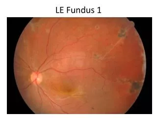
Intraretinal Hemorrhage HTN retinopathy with AV nicking and mild vascular tortuosity Mild NPDR Picture artifact. Large optic cup Prominent foveal reflex
· · Myopic degeneration with PPA and macular Chorioretinal atrophy Hypertensive retinopathy with Arteriolar attenuation. Increased arterial reflex. AV nicking. Tigroid Fundus. Large optic cup Proliferative retionpathy with regressed NVD. Previous PRP
Proliferative retinopathy with previous PRP Diabetic retinopathy with microaneurysms with macular exudates Nonexudative AMD with Drusen and RPE dropout Subfoveal CNVM with surrounding subretinal hemorrhage and exudates
Vitreous haze. Fleck Retinopathy chorioretinal scar with fibrovascular stalk secondary to focal chorioretinitis AMD with Mild intermediate drusen Advanced AMD with Geographic Atrophy involving fovea
POHS with regressed macular CNVM, PPA, and punched out CR scar AMD with severe large drusen Myopic Degeneration Flat choroidal Nevus adjacent to optic nerve
Diabetic retinopathy with Macular exudates at foveal edge, with possible CSME Diabetic retinopathy with Macular exudates POHS regressed macular CNVM with, PPA, and punchedout CR scar Conjunctival nevus