Understanding the Skeletal System: Structure, Function, and Types of Bone Tissue
The skeletal system forms the framework of the body, providing shape, support, and protection for vital organs. It consists of bones, cartilage, tendons, and ligaments. Key functions include support, protection, movement, storage of minerals like calcium and phosphorous, and blood cell production in bone marrow. Cartilage types include hyaline, elastic, and fibrocartilage, while bones are classified into axial and appendicular groups. Bone structure is complex, comprising various levels of composition and texture, which are essential for overall health and function.

Understanding the Skeletal System: Structure, Function, and Types of Bone Tissue
E N D
Presentation Transcript
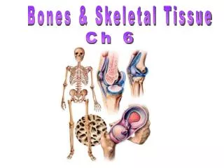
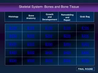
Skeletal System Bones and Bone Tissue
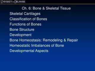
Skeletal System • Is the framework of the body • Provides shape to the body and protection for • organs and soft tissues • Consists of cartilage, bones, tendons and ligaments • Functions • Support:Bone is hard and rigid, bears body weight • Cartilage provides firm and flexible support, such as cartilage in nose, external ear, thoracic cage and trachea • Ligaments attach bone to bone & hold them together • Protection: Bones of skull protects brain • Ribs, sternum, vertebrae protect organs of thoracic cavity
Functions of Skeletal System • Movement: Skeletal muscles attach to bones by tendons • Contraction of skeletal muscles moves the bones, produce body movement • Storage:Ca and P. Stored then released as needed. Fat stored in marrow cavities • Blood cell production:Blood cells and platelets formation takes place in bone marrow of bones
Basic Structure of Skeletal Cartilage • Consists mostly of water – accounts for resilience • contains no nerves and blood vessels • Surrounded by double layer of dense irregular connective tissue – Perichondrium • Outer layer: Contains fibroblasts • Inner layer: More delicate, has fewer fibers, contains chondroblasts and chondrocytes • Blood vessels and nerves penetrate the outer layer of pericardium but do not enter cartilage matrix • Nutrients diffuse through matrix to reach chondrocytes Skeletal Cartilages
Skeletal Cartilages • Growth of Cartilage: • Cartilage grows in two ways: • Appositional growth – Add new matrix and chondrocytes to the outside of tissue • Interstitial growth –Chondrocytes within the tissue divide and add more matrix between the cells
Skeletal Cartilages • Types of Skeletal Cartilage: • Hyaline, Elastic and Fibrocartilage • Hyaline Cartilage: Contains fine collagen fibers in matrix, Include • Articular cartilages : cover the ends of bones at movable joints • Costal cartilages: connect the rib to sternum • Respiratory cartilages: forms skeleton of larynx • Nasal cartilages: support external nose • Elastic Cartilage: Contains collagen and elastic fibers - external ears and epiglottis • Fibrocartilage: Thick bundles of collagen fibers, compressible and tough • Found in Menisci and intervertebral discs
Classification of Bones • Human skeleton consists of 206 bones • And divided into two groups: • Axial Skeleton: • Consists of skull bones, vertebral column and rib cage • Protect and support body parts • Appendicular Skeletan: • Consists of bones of upper and lower limbs and girdles ( shoulder and hip bones) • Bones of limbs – help in movement
Classification of Bones • Bones are classified by their shape as long, short, flat and irregular • Long Bones: • Are long and thin • Are found in arms, legs, hands, feet, fingers, and toes • Flat Bones • Are thin, flattened shape, usually curved • Are found in the skull, sternum, ribs, and scapula
Classification of Bones • Irregular Bones • Have complex shapes • Examples: • spinal vertebrae • pelvic bones • Short Bones • Are small and thick • Examples: • ankle • wrist bones
Bone Structure • Three levels of structure: • Chemical • Gross • Microscopy
Chemical Composition of Bone • Consists of both organic and inorganic components • Organic components include: • Cells ( Osteoprogenitor cells, osteoblasts, osteocytes and osteoclasts) and Osteoid, the organic part of the matrix • Osteoid (35%) consists mainly of collagen and proteoglycans • Inorganic components • 65% of bone tissue is calcium phosphate crystal called hydroxyapatites, CaPO4 crystals
Gross Anatomy • Bone Markings • Most bones contain features on external surface • Depressions and openings along bone surface, passage for blood vessels and nerves • Projections where tendons and ligaments attach and at articulations with other bones • Include heads, trochanters, spines etc.
Bone textures • Bone tissue is classified as woven or lamellar bone based on collagen fibers organization within bone matrix • Woven bone. Collagen fibers randomly oriented. • First formed • During fetal development • During fracture repair • Then Woven bone is remodeled into lamellar bone • Lamellar bone • Mature bone, organized in sheets called lamellae. Collagen fibers are oriented in one direction in each layer, but in different directions in different layers for strength.
Bone textures • Bones, whether woven or lamellar can be classified • according to amount of bone matrix relative to amount of space • Compact bone: • Contains dense outer layer, less space • Cancellous or spongy bone: • Has less bone matrix & more space • Consists of interconnecting rods or plates of bones called trabeculae
Structure of Long Bone • Diaphysis • Shaft, long axis of bone • Made up of Compact bone • Surrounds central medullar or marrow activity • Red marrow - blood cell formation • Yellow marrow – adipose tissue • Epiphysis • End of the bone • Cancellous bone • Joint surface of epiphysis is covered with articular (hyaline) cartilage, cushions the bone ends • Epiphyseal plate:growth plate • Growth in length occurs at E. plate • Separates epiphysis from diaphysis • When bone stops growing in length becomes Epiphyseal line
Structure of Long Bone • Membranes • External surface of bone is covered by double layer membrane called Periosteum • Fibrous layer – Outer fibrous layer is dense irregular connective tissue contains blood vessels and nerves • Cellular layer – Inner single layer of bone cells consists of osteoblasts, osteoclasts, osteochondral progenitor cells • Periosteum is attached to underlying bone by Perforating or Sharpey`s fibers, made up of collagen • Periosteum provides anchoring points for tendons and ligaments
Structure of Long Bone • Membranes • Endosteum: • Single layer of cells that lines all internal spaces, such as medullar cavity • Contains osteoblasts, osteoclasts, osteochondral progenitor cells
Structure of Flat, Short, and Irregular Bones • Flat Bones • No diaphyses, epiphyses • Sandwich of cancellous between two layers of compact bone, eg. Parietal bone of skull • Short and Irregular Bone • Similar to structure of epiphyses of long bones • Compact bone that surrounds cancellous bone center with small spaces filled with marrow • Are not elongated and no diaphyses
Location of Hematopoietic Tissue in Bones • Hematopoietic tissue, red marrow is found in trabeculae of spongy bone of long bones, diploe of flat bones (sternum) and in some irregular bones (hip bones)
Microscopic Anatomy of Bone • Four major type of Bone cells • Osteoblasts • Osteocytes • Osteoclasts • Stem cells or osteochondral progenitor cells
Bone Cells • Osteoblasts • Immature bone cells that secrete organic components of matrix known as Osteoid • Osteoblasts surrounded by bone matrix, as the material calcifies, the cell is trapped in a space called a lacuna • And becomes osteocytes (mature bone cells)
Bone Cells • Osteocytes • Mature bone cells that maintain the bone matrix • Live in lacunae • Are between layers (lamellae) of matrix • Connect by cytoplasmic extensions through canaliculi in lamellae • Do not divide • Maintains protein and mineral content of matrix • Helps repair damaged bone
Bone Cells • Osteoclasts • Giant, mutlinucleate cells • Secrete acids and protein-digesting enzymes • Breakdown bone by dissolving bone matrix
Bone Cells • Osteoprogenitor Cells • Mesenchymal stem cells that divide to produce osteoblasts • Are located in inner layer of perichondrium, inner layer of periosteum and endosteum • Assist in fracture repair
Cancellous (Spongy) Bone • Consists of interconnecting rods or plates of bone called Trabeculae • No blood vessels in trabeculae • The space between trabeculae is filled with red bone marrow: • which has blood vessels • forms red blood cells • and supplies nutrients to osteocytes
Compact Bone • The basic unit of mature compact bone is Osteon or Haversian system • Osteon is a group of hollow tubes of bone matrix, one placed outside the next • Osteon consists of single central canal and around a canal contains blood vessels • Osteocytes are arranged in concentric lamellae
Compact Bone • Perforating (Volkmann`s )Canals • Perpendicular to the central canal • Carry blood vessels into bone and marrow • Circumferential Lamellae • Present on outer surface of compact bone • Binds osteons together • Interstitial Lamellae • Present in between osteons
Bone Development • Bone formation during fetal development occurs in two patterns: • Intramembranous ossification • Takes place in connective tissue membrane • Endochondral ossification • Takes place in cartilage • Both methods of ossification • Produce woven bone that is then remodeled • After remodeling, formation cannot be distinguished as one or other
Intramembranous Ossification • Takes place in fibrous connective tissue membrane formed from embryonic mesenchyme cells around the developing brain • Starts at 8th week & completes by age 2 • Forms many skull bones, part of mandible, diaphyses of clavicles • Mesenchyme cell in the membrane become osteochondral progenitor cell • Osteochondral progenitor cell forms osteoblast
Intramembranous Ossification • Osteoblast produce bone matrix and collagen fiber • And become osteocyte and develop trabeculae • More osteoblast gather around trabeculae and produce more bone • Trabeculae join together and form cancellous bone
Intramembranous Ossification • Cells in the spongy cell produce red bone marrow • Cells surrounding the developing bone forms periosteum • Osteoblasts from the periosteum on bone matrix produce compact bone
Endochondral Ossification • Bones of the base of the skull, part of the mandible, epiphyses of the clavicles, and most of remaining bones develop through endochondral ossification • Mesenchyme cells develop into chondroblasts which secrete the matrix of hyaline cartilage & surrounded by perichondrium except where joint formation takes place
Endochondral Ossification • Chondroblasts becomes chondrocytes; Chondrocytes in the center of hyaline cartilage: • Enlarge, calcify, and die, leaving cavities in cartilage • Blood vessels grow around the edges of the cartilage and osteochondral progenitor cells in the perichondrium change to osteoblasts • Perichondrium becomes periosteum when osteoblasts begin to form bone • Osteoblast produce compact bone on the surface of cartilage and forms the Bone collar
Endochondral Ossification • Blood vessels enter the cartilage • Connective tissue surrounding the blood vessels bring osteoblasts and osteoclasts • Osteoblasts secrete bone matrix and changes the calcified cartilage of diaphysis into cancellous bone • Bone formation area – Primary ossification center • Osteoclasts remove the bone from center and forms medullar cavity • Forms red bone marrow
Endochondral Ossification • Capillaries and osteoblasts enter the epiphyses creating secondary ossification centers • Epiphyses fill with spongy bone: • All the cartilage is replaced by bones except in epihhyseal plate and an articular sufaces • In mature bone, compact and cancellous bone are fully developed and epiphyseal plate becomes epiphyseal line
Bone Growth • Bones increase in size only by Appositional growth • Formation of new bone takes place on the surface of older bone or cartilage • Growth in Bone Length • Growth in bone length occurs at the epiphyseal plate • Growth at epiphyseal plate involves the formation of new cartilage by • Interstitial cartilage growth • Followed by Appositional bone growth on the surface of the cartilage
Growth in Bone Length • Epiphyseal plate is organized into 4 zones: • Zone of Resting Cartilage: • Nearest to epiphysis, contains randomly arranged chondrocytes • Do not involve in bone growth • Zone of Proliferation: • Contains actively dividing chondrocytes • Chondrocytes produce new cartilage through interstitial cartilage growth • As the cells divide, the epiphysis moves away from the diaphysis. This in turn produces length growth in bone
Growth in Bone Length • Zone of Hypertrophy: • Contains mature and enlarge • chondrocytes • Zone of Calcification: • Matrix is calcified, chondro- • cytes die • Calcified cartilage is • replaced by bone
Growth at Articular Cartilage • Growth at articular cartilage increases the epiphyses size • Also growth at articular cartilage increases size of bones with no epiphyses: e.g., short bones • Articular cartilage persists throughout life and does not ossified as epiphyseal plate
Growth in Bone Width • Bones increases in thickness or long bones increase • in width, because appositional bone growth takes place • beneath periosteum
Factors Affecting Bone Growth • Size and shape of a bone determined genetically but can be modified and influenced by nutrition and hormones • Nutrition • Lack of calcium, protein and other nutrients during growth and development can cause bones to be small • Vitamin D • Necessary for absorption of calcium from intestines • Can be eaten or manufactured in the body when skin is exposed to sunlight • Rickets: lack of vitamin D during childhood • Have bowed bones • Osteomalacia: lack of vitamin D during adulthood leading to softening of bones
Factors Affecting Bone Growth • Vitamin C • Necessary for collagen synthesis by osteoblasts • Scurvy: deficiency of vitamin C, causes hemorrhage because of lack of collagen deficiency • Lack of vitamin C also causes wounds not to heal, teeth to fall out
Bone Remodeling • Bone Remodeling: Old bone is replaced with new bone • Osteoclasts remove old bone and osteoblast forms new bone • Bone remodeling converts woven bone into lamellar bone • And involve in bone growth, changes in bone shape, adjustments in bone due to stress, bone repair, and Ca ion regulation • Caused by migration of Basic Multicellular Units • Groups of osteoclasts and osteoblasts that remodel bones • Bone constantly removed by osteoclasts and new bone formed by osteoblasts
Bone Repair • Bone undergo repair after damage • Has four major steps: • Hematoma formation: • Bone fracture damages blood vessels in bone & periosteum and hematoma forms • Hematoma - Localized mass of blood released from blood vessels, Clot formation stop the bleeding • Inflammation and swelling occurs after injury
Bone Repair • Callus formation. Callus is mass of tissue that forms at a fracture site and connects the broken ends of the bone • Internal Callus– Forms between the ends of broken bones • Several days after fracture blood vessels grow into clot • Macrophages clean up debris, • osteoclasts break down dead bone • tissue, fibroblasts produce collagen and helps in put the bone together • Osteoprogenitor cells from the periosteum of healthy bone tissue produce • Chondroblasts - which secrete cartilage tissue • Ostoeblasts- which secrete bone matrix • New bone is formed
Bone Repair • External Callus – Forms collar around opposite ends of bone fragments • Periosteal osteochondral progenitor cells osteoblasts and chondroblasts • Produce bones and cartilage
Bone Repair • Callusossification: • Callus replaced by woven, cancellous bone • Bone remodeling: Replacement of woven bone and damaged material by compact bone