Download
1 / 34
370 likes | 708 Vues
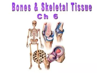
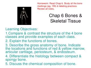
Homework: Read Chap 6. Study all the bone markings (pg. 109) & labeling practices well . Review all notes. Chap 6 Bones & Skeletal Tissue. Learning Objectives: 1.Compare & contrast the structure of the 4 bone classes and provide examples of each class. 2. Explain the functions of bones.

E N D
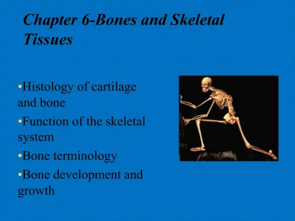
Homework: Read Chap 6. Study all the bone markings (pg. 109) & labeling practices well. Review all notes. Chap 6 Bones & Skeletal Tissue Learning Objectives: 1.Compare & contrast the structure of the 4 bone classes and provide examples of each class. 2. Explain the functions of bones. 3. Describe the gross anatomy of bone. Indicate the locations and functions of red & yellow marrow, articular cartilage, periosteum, & endosteum. 4. Differentiate the histology between compact & spongy bone. 5. Discuss the chemical composition of bone.
PREDICT • How many bones in the human skeleton?
Brainstorming Instructions: Working in small groups, without your book, name as many functions as you can in 2 minutes. Question: What are all the things that our skeleton (or bone) does for us? Note: There are at least 5 distinct things!
Functions of Bones • Support • Protection • Movement • Mineral storage • Blood cell formation
Review (Chap 4) 1. What kind of cartilage makes up the external ear? 2. What is the name of the most prominent kind of cartilage found in the costal areas (ribs), nose, shoulders, elbows, etc. 3. What is the name of the thick, pad-like cartilage of the knee and discs between the vertebrae?
How Are Bones Classified? Pg105 • The skeleton is divided into 2 main groups: a) _______ (skull, vertebrae & ribs) b) appendicular (limbs, shoulder, hip) areas. • From here, bones are further classified by their shape.
Shape - Long Bones Long bones – _________ than they are wide (e.g., humerus)
Shape - Short • Short bones • _____-shaped bones of the wrist and ankle • Bones that form within tendons (e.g., sesamoid bones such as the patella) patella
Shape - Flat • Flat bones – __, flattened, and a bit curved (e.g., sternum, and most skull bones)
Shape - Irregular • Irregular bones – bones with _________ shapes (e.g., vertebrae and hip bones)
Gross Anatomy of Bones pg. 107 • Rarely smooth • Have projections, depressions, and openings called _____ markings
Group Activity: Bone Markings Pgs, 107-114 Instructions: Work together in small groups of 3 to complete the information. Goal: To become more familiar with bone markings (projections, depressions, openings) Time Estimate: 15 minutes
Bones continued Learning Objectives continued: 6. Identify & explain the anatomy of a long bone; understand all associated terms (pg115) 7. Identify & explain the anatomy of a microscopic cross-section of bone; understand all associated terms (pg 116) 8. Discuss stress on bones & their response (page 116) 9. Explain the 6 common types of fractures (page 119) Homework: Finish reading Chapter 6. Review all diagrams, notes, class activities, practices, etc. *Be sure you know Table 6.1 Bone Markings BEFORE going into the next chapter.
Warm-Up Activity Instructions: Working individually (within Chap 6), use your textbook to locate the correct answers. Write just the letter of the answer) Answers: 1) q, 2) k, 3) g, 4) r, 5) b, 6) o, 7) a, 8) e, 9) c, 10) p, 11) m, 12) j, 13) h, 14) L, 15) f, 16) N, 17) d, 18) i
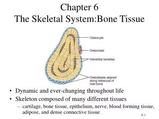
Bone Textures • Compact bone – _____ outer layer • Spongy bone – honeycomb of trabeculae filled with yellow bone marrow (internal to the compact bone)
New ‘Long Bone’ Vocabulary • Diaphysis • Medullary cavity • Epiphyses • Epiphyseal line • Periosteum • Osteoblasts • Osteoclasts • Sharpey’s fibers • Endosteum • Diploe • Red marrow Instructions: Define each term now in your notes (reference pages 160 – 161; also glossary in book may be used if appropriate)
Structure of Long Bones • Long bones consist of a ________ and an epiphysis • Diaphysis • Tubular shaft that forms the axis of long bones • Composed of compact bone that surrounds the medullary cavity • Yellow bone marrow (fat) is contained in the medullary cavity
Long Bone continued • Epiphyses • Expanded ____ of long bones • Exterior is compact bone, and the interior is spongy bone • Joint surface is covered with articular (hyaline) cartilage • Epiphyseal line separates the diaphysis from the epiphyses
Bone Membranes • Periosteum – double-layered protective membrane • Outer fibrous layer is dense regular connective tissue • Inner osteogenic layer is composed of osteoblasts and osteoclasts • Richly supplied with nerve fibers, blood, and lymphatic vessels, which enter the bone via nutrient foramina • Secured to underlying bone by Sharpey’s fibers (tufts of collagen fibers) • Endosteum – delicate membrane covering _______ surfaces of bone
Structure of Long Bone, pg. 115 Practice: Label your diagram.
Structure of Short, Irregular & Flat Bones • Thin plates of periosteum-covered compact bone on the outside with endosteum-covered spongy bone (diploë) on the inside • Have __ diaphysis or epiphyses • Contain bone marrow between the trabeculae
Where’s Red Marrow? • In infants • Found in the medullary cavity and all areas of spongy bone • In adults • Found in the _______ of flat bones, and the head of the femur and humerus
New Microscopic Bone Terminology Instructions: Define each term now in your notes. Use pages 116 – 117 or the glossary as appropriate. • Osteon or Haversian system • Lamella • Central (Haversian) canal • Perforating (Volkmann’s) canals • Lacunae • Canaliculi • Interstitial lamellae • Circumferential lamellae
Compact Bone (Microscopic View) • Haversian system or ________ – the structural unit of compact bone • Lamella – weight-bearing, column-like matrix tubes composed mainly of collagen • Haversian, or central canal – central channel containing blood vessels and nerves • Volkmann’s canals – channels lying at right angles to the central canal, connecting blood and nerve supply of the periosteum to that of the Haversian canal
Compact Bone – continued • Osteocytes – mature ______ ______ • Lacunae – small cavities in bone that contain osteocytes • Canaliculi – hairlike canals that connect lacunae to each other and the central canal
Compact Bone continued, pg 117 Label your practice diagram now. More About Bone Structure: http://youtube.com/watch?v=4qTiw8lyYbs
Bone Development • Osteogenesis and ossification – the process of bone tissue __________, which leads to: • The formation of the bony skeleton in embryos • Bone growth until early adulthood • Bone thickness, remodeling, and repair • Begins at week 8 of embryo development
Hormonal Regulation of Bone Growth During Youth • During _________ and childhood, epiphyseal plate activity is stimulated by growth hormone • During puberty, testosterone and estrogens: • Initially promote adolescent growth spurts • Cause masculinization and feminization of specific parts of the skeleton • Later induce epiphyseal plate closure, ending longitudinal bone growth
Bone Deposition & Mechanical Stress pg117 • Occurs where bone is injured or added strength is needed • __________ law – a bone grows or remodels in response to the forces or demands placed upon it • Trabeculae form along lines of stress • Large, bony projections occur where heavy, active muscles attach • Observations supporting Wolff’s law include • Long bones are thickest midway along the shaft (where bending stress is greatest) • Curved bones are thickest where they are most likely to buckle http://youtube.com/watch?v=qVougiCEgH8 About Bone Breakage & Repair:
Bone Fractures Pg.119-121 • Bone fractures are classified by: • The ________ of the bone ends after fracture • The completeness of the break • The orientation of the bone to the long axis • Whether or not the bones ends penetrate the skin
Activity: Bone Disorders Pg. 123 • Instructions: Work in groups of 3 - 4 • The various bone disorders are found on page 123. • The class will divide into groups. Groups will identify & discuss disorders (i.e., cause(s), symptoms, other pertinent information, etc.) • Disorders: 1. osteomalacia 2. rickets 3. osteoporosis 4. Paget’s disease Know the disorders for you next test! I may ask a question or two over these.