Bones and Skeletal Tissues
Bones and Skeletal Tissues. Structure and Function of Cartilage and Bone. Cartilage (review). Characteristics Resists tension AND compression Lots of collagen (strong) AND elastic fibers (flexible) High content of proteoglycans 80% water (resiliency/spring properties)


Bones and Skeletal Tissues
E N D
Presentation Transcript
Bones and Skeletal Tissues Structure and Function of Cartilage and Bone
Cartilage (review) Characteristics • Resists tension AND compression • Lots of collagen (strong) AND elastic fibers (flexible) • High content of proteoglycans 80% water (resiliency/spring properties) • No nerves or blood vessels • Chondroblasts make matrix until end of human adolescence • Mature Chondrocytes found in cavities called lacunae (pit) • Surrounded by the perichondrium (dense irregular connective tissue) that serves to: • Resists outward expansion • Nourish cartilage tissues
Hyaline Cartilage (review) • Looks glassy (hyalin = glass) • Few chondrocytes, all found in lacunae • Mostly matrix – lots of collagen
Hyaline Cartilage (add notes) Provides flexibility w/resilience Most abundant skeletal cartilage Locations: Articular – covers the ends of long bones, moving joings Costal – connects ribs (sternal) Respiratory –in larynx (voicebox), reinforces air passages of lungs Nasal – supports the nose
Elastic Cartilage (review) • Looks almost identical to hyaline BUT more elastic fibers more flexible! • Matrix appears more fibrous • More lacunae, closely spaced.
Elastic Cartilage (add) Elastic fibers allows for repeated “bending” Found in: the external ear Epiglottis (flap closes in larynx when we swallow)
Hyaline Cartilage Elastic Cartilage
Fibrocartilage (review) • Intermediate between dense regular CT and hyaline • Consists of rows of chondrocytes and collagen fibers • Compressible AND resists tension
Fibrocartilage (add) • Due to compressibility, found in areas that need padding due to stretch/pressure: • menisci (singular: meniscus) of the knee • intervertebral discs
Bones and Cartilages of the Human Body Figure 6.1
Growth of Cartilage Appositional – cells in the perichondrium secrete matrix at the surface of existing cartilage Grows “outward” Interstitial – lacunae-bound chondrocytes inside the cartilage divide and secrete new matrix, expanding the cartilage from within Grows from the inside of tissue • Area of appositional growth (black arrows) • Area of interstitial growth (white arrows)
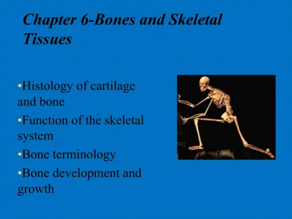
Function of Bones Support – “framework” that supports the body; cradles soft organs Protection –protective case for brain, spinal cord, vital organs Movement – levers for muscles Mineral storage – reservoir for minerals, especially calcium and phosphorus Blood cell formation – hematopoiesis occurs within the marrow cavities of bones
Bone Composition(organic) 4 cell types: genic Osteo cells • mitotic stem cells in membranes covering bones that create other bone cells • Osteoclasts: • Large cells that dissolve and break-down bone and its osteoid • Osteoblasts: • Young, bone cells that makes un-mineralized bone matrix, “osteoid” (ground substance, collagen, glycoproteins,etc.) • Osteocytes: • mature, bone cells that maintains osteoid
Bone Composition(inorganic) • Mineral Salts (Hydroxyapatites) • 65% bone by mass • mainly calcium phosphate, deposited around collagen of osteoid • responsible for hardness and compression resistance of bone • Deposited by osteoblasts, source of calcium and phosphate in blood plasma.
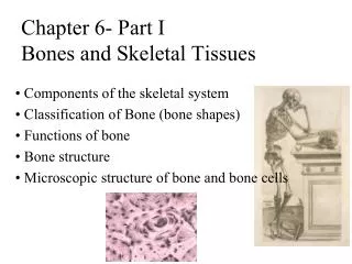
Skeletal Classification Axial skeleton – long axis of body bones of the skull, vertebral column, and rib cage Appendicular skeleton – bones of the limbs and girdle (shoulder, hip)
Classification of Bones: By Shape Long bones – longer than they are wide (ex. humerus) Named for relative length and width, not size (ex. fingers are also long bones!)
Short bones Cube-shaped bones of the wrist and ankle Bones that form within tendons (ex. patella) Classification of Bones: By Shape
Flat bones – thin, flattened, and a bit curved (ex. sternum, ribs, shoulder blades, skull) Classification of Bones: By Shape Figure 6.2c
Irregular bones –complicated shapes (ex. vertebrae and pelvis) Classification of Bones: By Shape
Bone Textures Compact bone – dense outer layer made up of osteons. Very supportive. Spongy bone – honeycomb of trabeculae filled with yellow marrow (fat) or red bone marrow (hematopoietic) • Trabeculae: • - needle-like extensions of bone • - disperse pressure without much weight • - resists compression on ends of bones • - marrow fills in between spaces
Membranes of Bones Porous with “nutrient foramens” to supply bone with nerves, lymph, and blood vessels Inner layer: osteogenic stem cells that differentiate (specialize) into bone cells like osteoblasts (bone forming) or osteoclasts (bone dissolving) cells. Periosteum: double-layered membrane on external surface of bones Outer layer: protective, fibrous dense irregular connective tissue Attach periosteum to bone Endosteum: delicate membrane covering internal surfaces of compact bone (next to marrow) or the trabeculae of spongy bones
Structure of Long Bone • Consists of a diaphysis (shaft) and an epiphyses (ends) • Diaphysis – • Forms the axis • Compact bone that surrounds hollow medullary cavity that contains yellow marrow (fat)
Structure of Long Bone • Epiphyses • compact bone exterior; spongy bone interior • joint surface covered w/ articular (hyaline) cartilage • cushions opposing bone ends during movement • Epiphyseal line, for bone lengthening, separates the diaphysis from the epiphyses
Structure of Short, Irregular, and Flat Bones Thin periosteum-covered compact bone on the outside with endosteum-covered spongy bone (diploë) on the inside Have no diaphysis or epiphyses Bone marrow fills spaces within trabeculae
Location of Hematopoietic Tissue (Red Marrow) In infants Found in the medullary cavity of long bones and all areas of spongy bone In adults Found in the diploë of flat bones, hip, and the head of the femur (thigh) and humerus (upper arm)
Micro-anatomy of Compact Bones Haversian system or Osteons: the structural unit of compact bone Volkmann’s canals: connects blood and nerve supply of the periosteum to the Haversian canal Haversian or Central Canal: central channel containing blood vessels and nerves Osteon periosteum Volkmann’s Canals
Lamella: an osteon’s weight-bearing, concentric matrix tubes (mostly of collagen) Micro-anatomy of Compact Bones Interstitial lamella: collagen matrix that fills in gaps between osteons • Circumferential lamella – extend under periosteum around the entire circumference of diaphysis
Micro-anatomy of Compact Bones • Osteoblasts: • Initially make matrix that hardens around them (lamella). • As they mature, osteoblasts become osteocytes. Osteocytes: trapped in cavities called lacunae,in between lamella Canaliculi: hairlike canals that connect lacunae to each other and the central canal