Bones and Skeletal Tissues
390 likes | 553 Vues
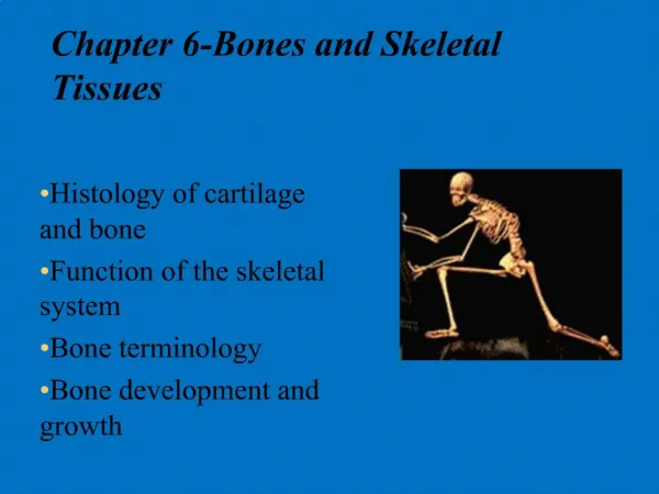
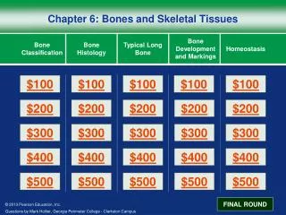
6. P A R T A. Bones and Skeletal Tissues. Function of Bones. Support – form the framework that supports the body and cradles soft organs Protection – provide a protective case for the brain, spinal cord, and vital organs Movement – provide levers for muscles. Function of Bones.

Bones and Skeletal Tissues
E N D
Presentation Transcript
6 P A R T A Bones and Skeletal Tissues
Function of Bones • Support – form the framework that supports the body and cradles soft organs • Protection – provide a protective case for the brain, spinal cord, and vital organs • Movement – provide levers for muscles
Function of Bones • Mineral storage – reservoir for minerals, especially calcium and phosphorus • Blood cell formation – hematopoiesis occurs within the marrow cavities of bones
Bones and Cartilages of the Human Body Figure 6.1
Classification of Bones: By Shape • Long bones – longer than they are wide (e.g., humerus) Figure 6.2a
Classification of Bones: By Shape • Short bones • Cube-shaped bones of the wrist and ankle • Bones that form within tendons (e.g., patella) Figure 6.2b
Classification of Bones: By Shape • Flat bones – thin, flattened, and a bit curved (e.g., sternum, and most skull bones) Figure 6.2c
Classification of Bones: By Shape • Irregular bones – bones with complicated shapes (e.g., vertebrae and hip bones) Figure 6.2d
Structure of Long Bone Figure 6.3a
Bone Markings • Bulges, depressions, and holes that serve as: • Sites of attachment for muscles, ligaments, and tendons • Joint surfaces • Conduits for blood vessels and nerves
Four Types of Bone Cells • Osteoprogenitor cells • stem cells • Osteoblasts • produce new bone • Eventually become osteocytes • Osteocytes • mature bone cells • Osteoclasts • are involved in bone resorption (breaking down & releasing things like calcium into the bloodstream)
Gross Anatomy of Bones: Bone Textures • Compact bone – dense outer layer • Spongy bone – honeycomb of trabeculae
Microscopic Structure of Bone: Compact Bone • Haversian system, or osteon – the structural unit of compact bone • Lamella – weight-bearing, column-like matrix tubes composed mainly of collagen • Haversian, or central canal – central channel containing blood vessels and nerves • Volkmann’s canals – channels lying at right angles to the central canal, connecting blood and nerve supply of the periosteum to that of the Haversian canal
Microscopic Structure of Bone: Compact Bone • Osteocytes – mature bone cells • Lacunae – small cavities in bone that contain osteocytes • Canaliculi – hairlike canals that connect lacunae to each other and the central canal
Ossification • Begins in the embryo and continues as the skeleton grows during childhood and adolescence. • Even after the adult bones have formed, ossification continues.
Intramembranous Ossification • From a connective tissue membrane into bone
Endochondral Ossification • Begins with a hyaline cartilage model.
Epiphyseal Line Formation • Endochondral ossification of a long bone occurs in progressive stages. • Bone growth is complete when each epiphyseal plate has ossified and the epiphyseal line has formed. • Depending on the bone, epiphyseal plate ossification occurs between the ages of 10 and 25 years.
Bone Growth • Interstitial growth occurs in the epiphyseal plate as chondrocytes undergo mitosis • Appositional growth occurs within the periosteum.
Bone Remodeling • Although adult bone size has been reached, the bone continues to reshape itself throughout a person’s lifetime in a constant process of bone resorption and deposition.
Bone Remodeling • The continual deposition of new bone tissue and the removal (resorption) of old bone tissue.
Types of Bone Fractures • Compound (open) – bone ends penetrate the skin • Impacted – one broken end driven into the other end of the bone • Pott’s – distal fibula • Colles’ – distal radius
Common Types of Fractures • Comminuted – bone fragments into three or more pieces; common in the elderly • Stress – broken but not displaced, not broken all the way through
Common Types of Fractures • Greenstick – incomplete fracture where one side of the bone breaks and the other side bends; common in children
Stages in the Healing of a Bone Fracture • Hematoma formation • A mass of clotted blood (hematoma) forms at the fracture site Figure 6.13.1
Stages in the Healing of a Bone Fracture • Fibrocartilaginous callus forms • forms a few days after the fracture Figure 6.13.2
Stages in the Healing of a Bone Fracture • Bony callus formation • New bone trabeculae appear in the fibrocartilaginous callus • Fibrocartilaginous callus converts into a bony (hard) callus • Bone callus begins 3-4 weeks after injury, and continues until firm union is formed 2-3 months later Figure 6.13.3
Stages in the Healing of a Bone Fracture • Bone remodeling • Excess material on the bone shaft exterior and in the medullary canal is removed • Compact bone is laid down to reconstruct shaft walls Figure 6.13.4
Effects of Hormones • Thyroid hormone stimulates bone growth. • Growth hormone and thyroid hormone regulate and maintain normal activity at the epiphyseal plates until puberty. • Calcitonin inhibits osteoclast activity.
Homeostatic Imbalances • Osteoporosis • Group of diseases in which bone reabsorption outpaces bone deposit • Spongy bone of the spine is most vulnerable • Occurs most often in postmenopausal women • Bones become so fragile that sneezing or stepping off a curb can cause fractures
Osteoporosis: Treatment • Calcium and vitamin D supplements • Increased weight-bearing exercise • Hormone (estrogen) replacement therapy (HRT) slows bone loss • Natural progesterone cream prompts new bone growth • Statins increase bone mineral density
Classification of Bones • Axial skeleton – bones of the skull, vertebral column, and rib cage • Appendicular skeleton – bones of the upper and lower limbs, shoulder, and hip