Bones and Skeletal Tissues
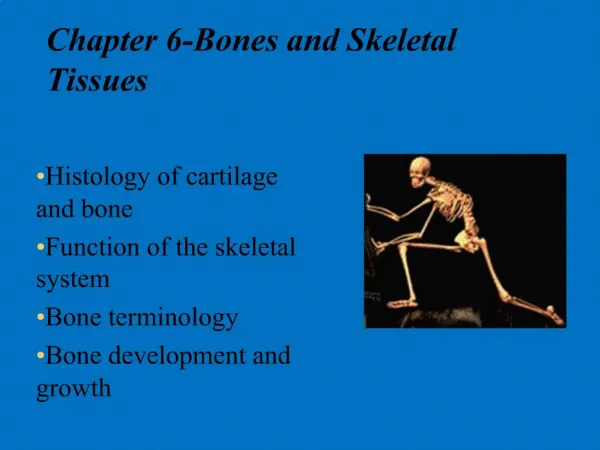
6. P A R T A. Bones and Skeletal Tissues. Bones and Cartilages of the Human Body. Figure 6.1. Classification of Bones. Axial skeleton – bones of the ___________________, ___________________, and ___________________


Bones and Skeletal Tissues
E N D
Presentation Transcript
6 P A R T A Bones and Skeletal Tissues
Bones and Cartilages of the Human Body Figure 6.1
Classification of Bones • Axial skeleton – bones of the ___________________, ___________________, and ___________________ • Appendicular skeleton – bones of the upper and lower ___________________, ___________________, and ___________________
Classification of Bones: By Shape • ________________bones – longer than they are wide • Example: • _________________ • __________________ • __________________ Figure 6.2a
Classification of Bones: By Shape • ________________ bones • Cube-shaped bones • _________________ • __________________ • __________________ Figure 6.2b
Classification of Bones: By Shape • _______________ bones – thin, flattened, and a bit curved • _________________ • __________________ • __________________ Figure 6.2c
Classification of Bones: By Shape • _______________bones – bones with complicated shapes • _________________ • __________________ • __________________ Figure 6.2d
Function of Bones • Support – form the framework that ____________________ the body and ____________________ soft organs • Protection – provide a protective case for the ____________________, ____________________, ____________________ • Movement – provide ____________________ for muscles
Function of Bones • Mineral storage – ____________________ for minerals, especially ____________________ and ____________________ • Blood cell formation – ____________________ occurs within the ____________________ cavities of bones
Bone Textures • Compact bone – dense ____________________ that looks smooth and solid to the naked eye • Spongy bone – ____________________which looks like a honeycomb of small pieces –____________________ • Spaces between trabeculae filled with ____________________
Structure of Long Bone Figure 6.3a
Structure of Long Bone Diaphysis: • Tubular shaft that forms the axis of long bones • Composed of ____________________ bone that surrounds the ____________________ (marrow) cavity- contains yellow bone marrow (fat)
Structure of Long Bone • Epiphyses: • Expanded ends of long bones • Exterior- ____________________ bone • Interior- ____________________ bone • Joint surface is covered with ____________________ (hyaline) cartilage • Epiphyseal line separates the diaphysis from the epiphyses
Structure of Long Bone Figure 6.3
Structure of Long Bone Figure 6.3b
Structure of Long Bone Figure 6.3c
Bone Membranes • ____________________– double-layered protective membrane on external surface of bone • Outer fibrous layer - dense regular ____________________ tissue • Inner layer- ____________________(“bone-making”) and ____________________ (“bone-breaking”) • Richly supplied with ____________________, ____________________, and lymphatic vessels, that enter the bone via nutrient foramina • Secured to underlying bone by Sharpey’s fibers
Bone Membranes • ____________________ – delicate membrane covering internal surfaces of bone
Structure of Short, Irregular, and Flat Bones • Thin plates of _____________________________ compact bone on the outside • ____________________ bone (diploë) on the inside • Have no ____________________ or ____________________ • Contain bone marrow between the ____________________
Structure of a Flat Bone Figure 6.4
Location of Hematopoietic Tissue (Red Marrow) • In infants • Found in the medullary cavity and all areas of ____________________ bone • In adults • Found in the diploë of flat bones, and the head of the ________________________________ and _____________________________________
Microscopic Structure of Bone: Compact Bone • Haversian system (osteon) – the structural unit of compact bone • ___________________________________ – weight-bearing, column-like matrix tubes composed mainly of collagen • _______________________________________, or central canal – central channel containing blood vessels and nerves
Microscopic Structure of Bone: Compact Bone • ____________________ – mature bone cells • ____________________ – small cavities in bone that contain osteocytes
Microscopic Structure of Bone: Compact Bone Figure 6.6a, b
Microscopic Structure of Bone: Compact Bone Figure 6.6a
Microscopic Structure of Bone: Compact Bone Figure 6.6b
Microscopic Structure of Bone: Compact Bone Figure 6.6c
Chemical Composition of Bone: Organic • ____________________– bone-forming cells • ____________________ – mature bone cells • ____________________ – large cells that resorb or break down bone matrix • ____________________ – ground substance and collagen fibers
Chemical Composition of Bone: Inorganic • Mineral salts • Sixty-five percent of bone by mass • Mainly calcium phosphates • Responsible for bone ____________________ and its resistance to ____________________
Bone Development • ____________________ __________________ • And ____________________________________ – the process of bone tissue formation, which leads to: • The formation of the bony skeleton in embryos • Bone growth until early adulthood • Bone thickness, remodeling, and repair
Formation of the Bony Skeleton • Begins at week 8 of embryo development • _____________________________ ossification – bone develops from a fibrous membrane • ____________________ _________ossification – bone forms by replacing hyaline cartilage
Intramembranous Ossification • Formation of most of the flat bones of the skull and the clavicles • Fibrous connective tissue membranes are formed by ____________________ _____________cells