Download
1 / 53
530 likes | 597 Vues
Learn about bone formation through ossification, stages of bone development, types of ossification, and the growth patterns of bones postnatally. Understand the key processes of osteoblasts, formation of woven and lamellar bone, and how long bones grow in length through functional zones.

E N D
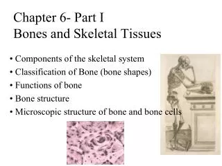
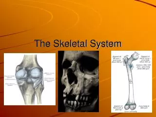
CHAPTER # 6(b) Bones & SKELETAL TISSUES
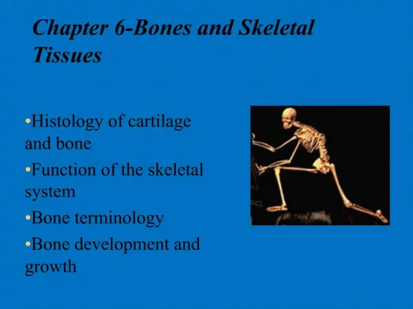
Bone Development • Osteogenesis (ossification)—bone tissue formation • Stages • Bone formation—begins in the 2nd month of development • Postnatal bone growth—until early adulthood • Bone remodeling and repair—lifelong
Two Types of Ossification • Intramembranous ossification • Membrane bone develops from fibrous membrane • Forms flat bones, e.g. clavicles and cranial bones • Endochondral ossification • Cartilage (endochondral) bone forms by replacing hyaline cartilage • Forms most of the rest of the skeleton
Mesenchymalcell Collagenfiber Ossificationcenter Osteoid Osteoblast 1 Ossification centers appear in the fibrousconnective tissue membrane. • Selected centrally located mesenchymal cells cluster and differentiate into osteoblasts, forming an ossification center. Figure 6.8, (1 of 4)
Osteoblast Osteoid Osteocyte Newly calcifiedbone matrix 2 Bone matrix (osteoid) is secreted within thefibrous membrane and calcifies.• Osteoblasts begin to secrete osteoid, which is calcified within a few days. • Trapped osteoblasts become osteocytes. Figure 6.8, (2 of 4)
Mesenchymecondensingto form theperiosteum Trabeculae ofwoven bone Blood vessel 3 Woven bone and periosteum form.• Accumulating osteoid is laid down between embryonic blood vessels in a random manner. The result is a network (instead of lamellae) of trabeculae called woven bone. • Vascularized mesenchyme condenses on the external face of the woven bone and becomes the periosteum. Figure 6.8, (3 of 4)
Fibrousperiosteum Osteoblast Plate ofcompact bone Diploë (spongybone) cavitiescontain redmarrow 4 Lamellar bone replaces woven bone, just deep tothe periosteum. Red marrow appears. • Trabeculae just deep to the periosteum thicken, and are later replaced with mature lamellar bone, forming compact bone plates. • Spongy bone (diploë), consisting of distinct trabeculae, per- sists internally and its vascular tissue becomes red marrow. Figure 6.8, (4 of 4)
Endochondral Ossification • Uses hyaline cartilage models • Requires breakdown of hyaline cartilage prior to ossification
Month 3 Birth Childhood toadolescence Week 9 Articularcartilage Secondaryossificationcenter Spongybone Epiphysealblood vessel Area ofdeterioratingcartilage matrix Epiphysealplatecartilage Hyalinecartilage Medullarycavity Spongyboneformation Bonecollar Bloodvessel ofperiostealbud Primaryossificationcenter 1 2 3 4 5 Bone collarforms aroundhyaline cartilagemodel. Cartilage in thecenter of thediaphysis calcifiesand then developscavities. The periostealbud inavades theinternal cavitiesand spongy bonebegins to form. The diaphysis elongatesand a medullary cavityforms as ossificationcontinues. Secondaryossification centers appearin the epiphyses inpreparation for stage 5. The epiphysesossify. Whencompleted, hyalinecartilage remains onlyin the epiphysealplates and articularcartilages. Figure 6.9
Week 9 Hyaline cartilage Bone collar Primaryossificationcenter 1 Bone collar forms aroundhyaline cartilage model. Figure 6.9, step 1
Area of deterioratingcartilage matrix 2 Cartilage in the centerof the diaphysis calcifiesand then develops cavities. Figure 6.9, step 2
Month 3 Spongyboneformation Bloodvessel ofperiostealbud 3 The periosteal bud inavadesthe internal cavities andspongy bone begins to form. Figure 6.9, step 3
Birth Epiphysealblood vessel Secondaryossificationcenter Medullarycavity 4 The diaphysis elongates and a medullary cavity formsas ossification continues. Secondary ossification centersappear in the epiphyses in preparation for stage 5. Figure 6.9, step 4
Childhood to adolescence Articular cartilage Spongy bone Epiphyseal platecartilage The epiphyses ossify. When completed, hyaline cartilageremains only in the epiphyseal plates and articular cartilages. 5 Figure 6.9, step 5
Month 3 Birth Childhood toadolescence Week 9 Articularcartilage Secondaryossificationcenter Spongybone Epiphysealblood vessel Area ofdeterioratingcartilage matrix Epiphysealplatecartilage Hyalinecartilage Medullarycavity Spongyboneformation Bonecollar Bloodvessel ofperiostealbud Primaryossificationcenter 1 2 3 4 5 Bone collarforms aroundhyaline cartilagemodel. Cartilage in thecenter of thediaphysis calcifiesand then developscavities. The periostealbud inavades theinternal cavitiesand spongy bonebegins to form. The diaphysis elongatesand a medullary cavityforms as ossificationcontinues. Secondaryossification centers appearin the epiphyses inpreparation for stage 5. The epiphysesossify. Whencompleted, hyalinecartilage remains onlyin the epiphysealplates and articularcartilages. Figure 6.9
Postnatal Bone Growth • Interstitial growth: • length of long bones • Appositional growth: • thickness and remodeling of all bones by osteoblasts and osteoclasts on bone surfaces
Growth in Length of Long Bones • Epiphyseal plate cartilage organizes into four important functional zones: • Proliferation (growth) • Hypertrophic • Calcification • Ossification (osteogenic)
Resting zone Proliferation zone Cartilage cells undergo mitosis. 1 Hypertrophic zone Older cartilage cells enlarge. 2 Calcification zone Matrix becomes calcified; cartilage cells die; matrix begins deteriorating. 3 Calcified cartilage spicule Osteoblast depositing bone matrix Ossification zone New bone formation is occurring. 4 Osseous tissue (bone) covering cartilage spicules Figure 6.10
Hormonal Regulation of Bone Growth • Growth hormone stimulates epiphyseal plate activity • Thyroid hormone modulates activity of growth hormone • Testosterone and estrogens (at puberty) • Promote adolescent growth spurts • End growth by inducing epiphyseal plate closure
Bone remodeling Bone growth Articular cartilage Cartilage grows here. Epiphyseal plate Cartilage is replaced by bone here. Bone is resorbed here. Cartilage grows here. Bone is added by appositional growth here. Cartilage is replaced by bone here. Bone is resorbed here. Figure 6.11
Bone Deposit • Occurs where bone is injured or added strength is needed • Requires a diet rich in protein; vitamins C, D, and A; calcium; phosphorus; magnesium; and manganese
Bone Deposit • Sites of new matrix deposit are revealedby the • Osteoid seam • Unmineralized band of matrix • Calcification front • The abrupt transition zone between the osteoid seam and the older mineralized bone
Bone Resorption • Osteoclasts secrete • Lysosomal enzymes (digest organic matrix) • Acids (convert calcium salts into soluble forms) • Dissolved matrix is transcytosed across osteoclast, enters interstitial fluid and then blood
Control of Remodeling • What controls continual remodeling of bone? • Hormonal mechanisms that maintain calcium homeostasis in the blood • Mechanical and gravitational forces
Hormonal Control of Blood Ca2+ • Calcium is necessary for • Transmission of nerve impulses • Muscle contraction • Blood coagulation • Secretion by glands and nerve cells • Cell division
Hormonal Control of Blood Ca2+ • Primarily controlled by parathyroid hormone (PTH) Blood Ca2+ levels Parathyroid glands release PTH PTH stimulates osteoclasts to degrade bone matrix and release Ca2+ Blood Ca2+ levels
Calcium homeostasis of blood: 9–11 mg/100 ml BALANCE BALANCE Stimulus Falling blood Ca2+ levels Thyroid gland Osteoclasts degrade bone matrix and release Ca2+ into blood. Parathyroid glands Parathyroid glands release parathyroid hormone (PTH). PTH Figure 6.12
Hormonal Control of Blood Ca2+ • May be affected to a lesser extent by calcitonin Blood Ca2+ levels Parafollicular cells of thyroid release calcitonin Osteoblasts deposit calcium salts Blood Ca2+ levels • Leptin has also been shown to influence bone density by inhibiting osteoblasts
Response to Mechanical Stress • Wolff’s law: A bone grows or remodels in response to forces or demands placed upon it • Observations supporting Wolff’s law: • Handedness (right or left handed) results in bone of one upper limb being thicker and stronger • Curved bones are thickest where they are most likely to buckle • Trabeculae form along lines of stress • Large, bony projections occur where heavy, active muscles attach
Load here (body weight) Head of femur Tension here Compression here Point of no stress Figure 6.13
Classification of Bone Fractures • Bone fractures may be classified by four “either/or” classifications: • Position of bone ends after fracture: • Nondisplaced—ends retain normal position • Displaced—ends out of normal alignment • Completeness of the break • Complete—broken all the way through • Incomplete—not broken all the way through
Classification of Bone Fractures • Orientation of the break to the long axis of the bone: • Linear—parallel to long axis of the bone • Transverse—perpendicular to long axis of the bone • Whether or not the bone ends penetrate the skin: • Compound (open)—bone ends penetrate the skin • Simple (closed)—bone ends do not penetrate the skin
Common Types of Fractures • All fractures can be described in terms of • Location • External appearance • Nature of the break
Stages in the Healing of a Bone Fracture • Hematoma forms • Torn blood vessels hemorrhage • Clot (hematoma) forms • Site becomes swollen, painful, and inflamed
Hematoma 1 A hematoma forms. Figure 6.15, step 1
Stages in the Healing of a Bone Fracture • Fibrocartilaginous callus forms • Phagocytic cells clear debris • Osteoblasts begin forming spongy bone within 1 week • Fibroblasts secrete collagen fibers to connect bone ends • Mass of repair tissue now called fibrocartilaginous callus
Externalcallus Internalcallus(fibroustissue andcartilage) Newbloodvessels Spongybonetrabecula 2 Fibrocartilaginouscallus forms. Figure 6.15, step 2
Stages in the Healing of a Bone Fracture • Bony callus formation • New trabeculae form a bony (hard) callus • Bony callus formation continues until firm union is formed in ~2 months
Bonycallus ofspongybone 3 Bony callus forms. Figure 6.15, step 3
Stages in the Healing of a Bone Fracture • Bone remodeling • In response to mechanical stressors over several months • Final structure resembles original
Healedfracture 4 Bone remodelingoccurs. Figure 6.15, step 4
Hematoma Externalcallus Bonycallus ofspongybone Internalcallus(fibroustissue andcartilage) Healedfracture Newbloodvessels Spongybonetrabecula 1 2 3 4 A hematoma forms. Fibrocartilaginouscallus forms. Bony callus forms. Boneremodelingoccurs. Figure 6.15
Homeostatic Imbalances • Osteomalacia and rickets • Calcium salts not deposited • Rickets (childhood disease) causes bowed legs and other bone deformities • Cause: vitamin D deficiency or insufficient dietary calcium
Homeostatic Imbalances • Osteoporosis • Loss of bone mass—bone resorption outpaces deposit • Spongy bone of spine and neck of femur become most susceptible to fracture • Risk factors • Lack of estrogen, calcium or vitamin D; petite body form; immobility; low levels of TSH; diabetes mellitus
Osteoporosis: Treatment and Prevention • Calcium, vitamin D, and fluoride supplements • Weight-bearing exercise throughout life • Hormone (estrogen) replacement therapy (HRT) slows bone loss • Some drugs (Fosamax, SERMs, statins) increase bone mineral density
Paget’s Disease • Excessive and haphazard bone formation and breakdown, usually in spine, pelvis, femur, or skull • Pagetic bone has very high ratio of spongy to compact bone and reduced mineralization • Unknown cause (possibly viral) • Treatment includes calcitonin and biphosphonates