Download
1 / 70
770 likes | 1.19k Vues
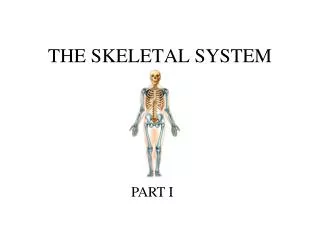
THE SKELETAL SYSTEM CHAPTERS 6 & 7. The Skeletal System. Parts of the skeletal system Bones (skeleton) Joints Cartilages Ligaments Divided into two divisions Axial skeleton Appendicular skeleton. Bones of the Human Body. The adult skeleton has 206 bones Two basic types of bone tissue

E N D
The Skeletal System • Parts of the skeletal system • Bones (skeleton) • Joints • Cartilages • Ligaments • Divided into two divisions • Axial skeleton • Appendicular skeleton
Bones of the Human Body • The adult skeleton has 206 bones • Two basic types of bone tissue • Compact bone • Homogeneous • Spongy bone • Small needle-like pieces of bone • Many open spaces Figure 5.2b
Functions of Bones • Support of the body • Protection of soft organs • Movement due to attached skeletal muscles • Storage of minerals • Blood cell formation • Storage of fats
Classification of Bones on the Basis of Shape – pg 70 SG Figure 5.1
Classification of Bones • Long bones • Typically longer than wide • Have a shaft with heads at both ends • Contain mostly compact bone • Examples: Femur, Humerus
Classification of Bones • Short bones • Generally cube-shape • Contain mostly spongy bone • Examples: Carpals, tarsals
Flat bones Thin and flattened Usually curved Thin layers of compact bone around a layer of spongy bone Examples: Skull, ribs, sternum Classification of Bones
Irregular bones Irregular shape Do not fit into other bone classification categories Example: Vertebrae and hip Classification of Bones
Epiphysis Diaphysis Articular cartilage Epiphyseal line Spongy bone Compact bone Medullary cavity Periosteum Gross Anatomy of the typical long bone – pg 72 of SG
Mature bone cells are Osteocytes Components
Haversian canal Lacunae Osteocytes Lamellae Canaliculi Volkmann’s canals Matrix Pg. text, 176 #A5 SG Microscopic Structure of Bone
Bone Markings • Surface features of bones • Sites of attachments for muscles, tendons, and ligaments • Passages for nerves and blood vessels • Categories of bone markings • Projections and processes – grow out from the bone surface • Depressions or cavities – indentations
Bone Markings Projections/sites of muscle and ligament attachment Projections/sites that form joints Head Facet Condyle Ramus • Tuberosity • Crest • Trochanter • Line • Tubercle • Epicondyle • Spine • Process
More Bone Markings Cavities Depressions/Openings allow blood vessels and nerves to pass Meatus Fossa Groove Fissure Foramen • Sinus
Table 7.2 pg 198 • Head – rounded articular process at the proximal end of a bone • Condyle – rounded articular process at the distal end of a bone • Epicondyle – a small raised area above a condyle for joint capsule attachment • Foramen – a short passageway through bone for vessels and nerves • Meatus – a long canal like passageway
Fossa – a depression in bone • Sinus – a cavity in bone lined by a mucous membrane • Trochanter – very large projection • Tuberosity – a large rounded projection for muscle attachment • Tubercle – a small rounded projection • Fissure – a slit like opening through bone • Facet – smooth flat articular surface
Crest – prominent ridge or elongated projection • Sulcus – furrow along a bone surface where a blood vessel, nerve or tendon is located • Spine – sharp, slender often pointed projection • Using an Anatomy Atlas, see if you can identify bone surface markings on the skeleton and unarticulated bones at the front of the room
Color Axial Skeleton Appendicular Skeleton With a key Pg 73 SG
The Axial Skeleton • Forms the longitudinal part of the body • Divided into three parts • Skull • Vertebral column • Bony thorax
Text pg. 199 – 204 Color each of the bones of the skull The Skull
The Skull • Two sets of bones • Cranium • Facial bones • Bones are joined by sutures • Only the mandible is attached by a freely movable joint
Paranasal sinuses Text pg. 211 • Paranasal sinuses
Text pg. 213 The Vertebral Column
Spinal abnormalities Lordosis Kyphosis Scoliosis Text pg. 226
Structure of a Typical Vertebrae Figure 5.16
Regional Characteristics Figure 5.17a–b
Regional Characteristics Figure 5.17c–d
Text pg. 217 The Vertebrae
Bony thorax Rib cage Text pg. 223
Forms a cage to protect organs
Made up of 3 parts Sternum Ribs Thoracic vertebrae
Pectoral girdle Text pg. 232 The Appendicular Skeleton
The Pectoral (Shoulder) Girdle • Composed of two bones • Clavicle – collarbone • Scapula – shoulder blade • These bones allow the upper limb to have exceptionally free movement