Cardiac Cycle: diastole Phase
240 likes | 597 Vues
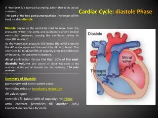
Cardiac Cycle: diastole Phase. A heartbeat is a two-part pumping action that takes about a second. This part of the two-part pumping phase (the longer of the two) is called diastole .

Cardiac Cycle: diastole Phase
E N D
Presentation Transcript
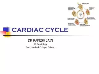
Cardiac Cycle: diastole Phase A heartbeat is a two-part pumping action that takes about a second. This part of the two-part pumping phase (the longer of the two) is called diastole. Diastole begins as the ventricles start to relax. Soon the pressures within the aorta and pulmonary artery exceed ventricular pressures, causing the semilunar valves to close (B2 murmur). As the ventricular pressure falls below the atrial pressure the AV valves open and the ventricles fill with blood. The ventricles fill to about 80% of capacity prior to contraction of the atria, the last event in diastole. Atrial contraction forces the final 20% of the end-diastolic volume(the volume of blood that exists in the ventricles at the end of diastole) into the ventricles. / SA nodecontracts Summary of Diastole: pulmonary and aortic valves close Ventricles relax => isovolumic relaxation AV valves open ventricles fill (about 80% of capacity) => inflow atria contract (ventricles fill another 20%) Contraction reaches AV node…
CardiacCycle: Systole Phase The second part of the pumping phase begins when the ventricles are full of blood. The electrical signals from the SA node travel along a pathway of cells to the ventricles, causing them to contract. This is called systole. As the ventricles start to contract, the ventricular pressure soon exceeds the atrial pressure, causing the AV valves to close (B1 murmur). As the ventricles continue to contract, the ventricular pressure exceeds the arterial pressures causing the semilunar valves open. Blood is forcefully ejected out of the ventricles and into the aorta and pulmonary artery. Summary of Systole: AV valves close ventricles contract => isovolumic contraction aortic and pulmonary valves open blood is ejected => ejection phase atria relax and fill with blood
Plan: Evaluation of the cardiac performance • On the cellular « scale » • muscularcells and cardiacmyocytes • myofibril / « Sarcomere » • proteins: actin / myosin • role of Ca++ • => « cell’s performance » • Laws and principles of haemodynamic: • Poiseuille or Darcy • Starling : preload and post load • «heart’s performance » • On the scale of the organ: the heart • Cardiac cycle • Relation pressure-volume • The regulation of the cardiac output
Physiology (1) All muscles derive from paraxial mesoderm. The three types of muscle (skeletal, cardiac and smooth) have significant differences. However, all three use the movement of actin against myosin to create contraction. Cardiac muscle is a type of involuntary striated muscles found in the walls of the heart, specifically the myocardium. Cardiac and smooth muscle contractions are stimulated by internal pacemaker cells which regularly contract, and propagate contractions to other muscle cells they are in contact with. Muscle is mainly composed of muscle cells. Within the cells are myofibrils; myofibrils contain sarcomeres, which are composed of actin and myosin. muscle cell or
1:Axon / 2:neuromuscularjunction / 3:muscle cell/ 4:myofibril Physiology (2) Myofibrils are cylindrical organelles. They are found within muscle cells or fibers. They are bundles of actomyosin filaments that run from one end of the cell to the other and are attached to the cell surface membrane at each end. The filaments of myofibrils, or myofilaments, consist of two types, thick and thin. Thin filaments consist primarily of the protein actin, coiled with nebulin filaments. Thick filaments consist primarily of the protein myosin, held in place by titin filaments. The filaments are organized into repeated subunits along the length of the myofibril. These subunits are called sarcomeres. The sarcomere is the “functional” basic unit of contraction.
sarcomere sarcomere sarcomere
Physiology (3) Sliding filament model of muscle contraction myosin relaxed Cardiac muscle requires extracellular calcium ions for contraction to occur. Like skeletal muscle, the initiation and upshoot of the action potential in ventricular muscle cells is derived from the entry of sodium ions across the sarcolemma in a regenerative process. Once the intracellular concentration of calcium increases, calcium ions bind to the protein troponin, which initiates contraction by allowing the contractile proteins, myosin and actin to associate through cross-bridge formation. actin filament contracted Shortening of 1 µm / sarcomere If 10 5sarcolemma (striated muscle) => Shortening of 10cm
Neuromuscularjunctionfor the nextpresentation… In contrast to skeletall muscle, cardiac muscle requires extracellular calcium ions for contraction to occur. Like skeletal muscle, the initiation and upshoot of the action potential in ventricular muscle cells is derived from the entry of sodium ions across the sarcolemma in a regenerative process. However, an inward flux of extracellular calcium ions through type calcium channels sustains the depolarization of cardiac muscle cells for a longer duration. Once the intracellular concentration of calcium increases, calcium ions bind to the protein troponin, which initiates contraction by allowing the contractile proteins, myosin and actin to associate through cross-bridge formation.
Poiseuille or Darcy’slaw/ (Ohm) • ∆P = Q x Rv / (Flow = Pressure/Resistance ) • ∆P => Mean gradient pressure • ∆Ps = PAo – POD • ∆Pp = PAP – PVP • Qs = SV x Hr(SV=> Stroke Volume = EDV – ESV ) Afterbirth: « serial circulation » without shunt => Qs=Qp => pressures in aorta and PA depend on Resistances Beforebirth: « parallel circulation » With shunts => Pao = PAP / Qs ≠ Qp Rp are high => Qpislow Rs are low (placenta) => Qsishigh
Frank-Starlinglaw • The Frank-Starling law of the heart states that the greater the volume of blood entering the heart during diastole (end-diastolic volume), the greater the volume of blood ejected during systolic contraction (stroke volume). • This allows the cardiac output to be synchronized with the venous return, arterial blood supply and humeral length[1] without depending upon external regulation to make alterations. • As the heart fills with more blood than usual, the force of the muscular contractions will increase. • The stretching of the muscle fibresincreases the affinity of troponin C for calcium, causing a greater number of cross-bridges to form within the muscle fibers; this increases the contractile force of the cardiac muscle. • The force that any single muscle fiber generates is proportional to the initial sarcomere length (known as preload), and the stretch on the individual fibers is related to the end-diastolic volume of the ventricle.
TA Toum
TA Toum
Parameter Values • End-diastolic volume (EDV) 120 ml • End-systolic volume (ESV) 50 ml • Stroke volume (SV) 70 ml • Ejection fraction (Ef) 58% • Heart rate (HR) 70 bpm • Cardiacoutput (CO) 4.9 L/mn • Cardiac index (CI) 2,5 -3,5 l/mn/m2 290 l / hour 7 056 l / day 2 575 440 l / year 180 280 800 l / 70 years
End systolic volume (afterload volume) Pressure mm Hg Starlinglaw Contractility 140 Systolic phase Ejection phase Closure of the aortic valves 80 Opening of the aortic valves SV (35ml) Isometric relaxation Isometric contraction Compliance 30 Opening of the AV valves 10 Closure of the AV valves Filling Volume ml 10 50 40 100 End diastolic volume (preload volume) Diastolic phase
Regulation of cardiac output the rate of contraction can be changed by nervous or hormonal influences, exercise and emotions. For example, the sympathetic nerves to heart accelerate heart rate and the vagus nerve decelerates heart rate. Qs = SV x Hr (SV=> Stroke Volume = EDV – ESV )
Evaluation of and for the « teacher » ! • How do youdefine a muscularfiberand a sarcomere ? • Explain the mechanism of shortening of a muscle fiber? • Whatis the Starling’slaw? • Whathappensduring the isovolumic relaxation concerning the heart’svalves ? • Define the « compliance » of the heart • How manylitersdoesthe heartpumpduring one year ? (bonus)