Download

1 / 89
940 likes | 1.3k Vues
Understand the types of sensory receptors, general senses, hearing, vision, and pain pathways in the human body. Learn about sensory transduction, receptor classifications, and pain perceptions. Dive into the complexities of somesthetic projection pathways and the fascinating world of chemoreceptors. Explore how the brain processes stimuli and reacts to pain signals.

E N D
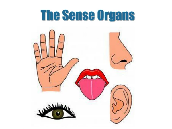
Chapter 16Sense Organs • Types of sensory receptors • General senses • Chemical senses • Hearing and equilibrium • Anatomy of the ear • Vision
Properties of Receptors • Receptor is any structure specialized to detect a stimulus (simple nerve ending or complex sense organ) • Sensory receptors are transducers converting stimulus energy into electrochemical energy = sensory transduction • Information about stimulus that can be conveyed • modality or type of stimulus or sensation • location of stimuli • each sensory receptor receives input from its receptive field • brain identifies site of stimulation = sensory projection • intensity (frequency, numbers of fiber & which fibers) • duration = change in firing frequency over time • phasic receptor - burst of activity & quickly adapt (smell & hair receptors) • tonic receptor - adapt slowly, generate impulses continually (proprioceptor)
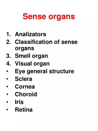
Classification of Receptors • By modality: • chemoreceptors, thermoreceptors, nociceptors, mechanoreceptors and photoreceptors • By origin of stimuli • interoceptors = detect internal stimuli • proprioceptors = sense position & movements of body • exteroceptors = sense stimuli external to body • By distribution • general (somesthetic) sense --- widely distributed • special senses --- limited to head
The General SensesUnencapsulated Nerve Endings • Found as receptors for the general senses • Dendrites not wrapped in connective tissue • Free nerve endings include • warm, cold & pain • Tactile discs are associated with cells at base epidermis • Hair receptors monitor the movement of hairs
Encapsulated Nerve Endings • Dendrites wrapped by glial cells or connective tissue • tactile (meissner) corpuscles • light touch & texture • krause end bulb • tactile corpuscles in mucous membranes • lamellated (pacinian) corpuscles • deep pressure, stretch, tickle & vibration • ruffini corpuscles • heavy touch, pressure, joint movements & skin stretching
Somesthetic Projection Pathways • First-order neuron or afferent neuron • from below head, enter the dorsal horn of spinal cord via spinal nerves • from head, enter pons and medulla from cranial nerve • touch, pressure & proprioception are carried on large, fast, myelinated axons • heat & cold are carried on small, unmyelinated, slow fibers • Second-order neuron • decussation of signals to opposite side in spinal cord or medulla • end in thalamus, except for proprioception (cerebellum) • Third-order neuron • extend from thalamus to primary somesthetic cortex of cerebrum on contralateral side
Pain • Nociceptors make us conscious of tissue injuries • forces us to care for minor injuries to prevent serious problems • Found in all tissues except the brain • Fast pain travels in myelinated fibers at 30 m/sec • sharp, localized, stabbing pain perceived with injury • Slow pain travels unmyelinated fibers at 2 m/sec • longer-lasting, dull, diffuse feeling • Somatic pain arises from skin, muscles & joints • Visceral pain from stretch, chemical irritants or ischemia of viscera (poorly localized) • Injured tissues release chemicals that stimulate pain fibers (bradykinin, histamine, prostaglandin)
Projection Pathway for Pain • General pathway • first-order neuron cell bodies in dorsal root ganglion of spinal nerves or trigeminal ganglion • second-order neurons decussate to other side & send fibers up spinothalamic tract to thalamus • gracile fasciculus carries visceral pain signals • third-order neurons reach primary somesthetic cortex (postcentral gyrus) • Spinoreticular tract • pain signals reach reticular formation, hypothalamus & limbic • trigger emotional and behavioral reactions • Referred pain is misinterpreted pain • brain “assumes” pain is coming from skin or superficial sites • heart pain felt in shoulder or arm because both send pain input to spinal cord segments T1 to T5
CNS Modulation of Pain • Intensity of pain is affected by state of mind • Endogenous opiods (enkephalins, endorphins 7 dynorphins) • produced by CNS and other organs under stress • found especially in dorsal horn of spinal cord, explaining the spinal gating of pain • act as neuromodulators blocking the transmission of pain • Spinal gating stops pain signals at dorsal horn • descending analgesic fibers from reticular formation travel down reticulospinal tract to dorsal horn • secrete inhibitory substances that block pain fibers from secreting substance P • pain signals never ascend • dorsal horn fibers inhibited by input from mechanoreceptors (rubbing a sore arm reduces pain)
Spinal Gating of Pain Signals Normal pain pathways(red arrows) is inhibited by many mechanisms.
The Chemical Sense -- Taste • Gustation is the sensation of taste resulting from the action of chemicals on the taste buds • Lingual papillae • filiform (no taste buds) • important for texture • foliate (no taste buds) • fungiform • at tips & sides of tongue • vallate (circumvallate) • at rear of tongue • contains 1/2 of taste buds
Taste Bud Structure • Cell group = taste cells, supporting cells, and basal cells • taste cells with a apical microvilli serving as a receptor surface • taste cells synapse with sensory nerve fibers at their base
Physiology of Taste • To be tasted, molecules must dissolve in saliva • 5 primary sensations: salty, sweet, sour, bitter & umami (taste of amino acids such as MSG) • taste is also influenced by food texture, aroma, temperature, and appearance • mouthfeel is detected by lingual nerve branches in papillae • hot pepper stimulates free nerve endings (pain) • Sweet tastes concentrated on tip of tongue, salty & sour on lateral margins of tongue & bitter at rear • all tastes can be detected throughout the tongue surface • Mechanisms of action • sugars, alkaloids & glutamates bind to receptors & activate 2nd messenger systems • sodium & acids penetrate cells & depolarize them directly
Projection Pathways for Taste • Innervation of the taste buds • facial nerve (VII) for the anterior 2/3’s of the tongue • glossopharyngeal nerve (IX) for the posterior 1/3 • vagus nerve (X) for palate, pharynx & epiglottis • All fibers project to solitary nucleus in medulla • Cells project to hypothalamus & amygdala • activate autonomic reflexes such as salivation, gagging & vomiting • Cells project to thalamus & then postcentral gyrus of the cerebrum • conscious sense of taste
The Chemical Sense -- Smell • Receptor cells for olfaction form olfactory mucosa • smell is highly sensitive (more so in women than men) • distinguish as many as 10,000 odors • Covers 5cm2 of superior concha & nasal septum
Olfactory Epithelial Cells • Olfactory cells • neurons with 20 cilia called olfactory hairs • binding sites for odor molecules in thin layer of mucus • axons pass through cribriform plate • survive 60 days • Supporting cells • Basal cells divide
Physiology of Smell • Odor molecules must be volatile • bind to a receptor on an olfactory hair triggering the production of a second messenger • opens the ion channels & creates a receptor potential • Receptors adapt quickly due to synaptic inhibition in the olfactory bulbs • Bulb cells form the axons of the olfactory tracts • lead to temporal lobe, amygdala, hypothalamus • emotional responses to odors • cough, salivate, sneeze or vomit in response to odors • cerebral cortex sends feedback to bulb cells • changing quality & significance of odors when hungry
The Nature of Sound • Sound is any audible vibration of molecules • Vibrating object pushes air molecules into eardrum making it vibrate Molecules collide with eardrum & make it vibrate.
Prolonged Sounds 90 dB can cause damage. Pitch and Loudness • The frequency at which parts of the ear vibrate give us sense of pitch (high or low pitched sounds) • hearing range is 20 - 20,000 Hz (cycles/sec) • speech is within 1500-4000 where hearing is most sensitive • Loudness is perception of intensity of sound energy • how much the air molecules are compressed in decibels (dB)
Outer Ear • Fleshy auricle (pinna) directing air vibrations down auditory canal (external auditory meatus) • cartilagenous & bony, S-shaped tunnel ending at eardrum
Middle Ear • Air-filled cavity in temporal bone separated from air outside the head by tympanic membrane • 1 cm in diameter, slightly concave, freely vibrating membrane • Tympanic cavity continuous with mastoid air cells • Tympanic cavity filled with air by auditory tube (eustachian tube) connected to nasopharynx • opens during swallowing or yawning to equalize air pressure on both sides of eardrum • Ear ossicles span tympanic cavity • malleus attached to eardrum, incus, stapes attached to membranous oval window of inner ear • stapedius & tensor tympani muscles attach to ossicles
Anatomy of Middle Ear • Middle ear is cavity containing ear ossicles.
vestibular apparatus cochlea Inner Ear • Passageways in temporal bone = bony labyrinth • Fleshy tubes lining bony tunnels = membranous labyrinth • filled with endolymph (similar to intracellular fluid) • floating in perilymph (similar to cerebrospinal fluid)
Anatomy of the Cochlea 2.5 coils • Stereocilia of hair cells attached to gelatinous tectorial membrane. • Hearing comes from inner hair cells -- outer ones adjust cochlear responses to different frequencies increasing precision 3 fluid-filled chambers Organ of Corti
Physiology of Hearing -- Middle Ear • Eardrum vibrates quite easily • 18 times the area of the oval window • creates enough force/unit area at oval window to vibrate the endolymph in the scala vestibuli • Protection of cochlea by muscle contraction in response to loud noises (tympanic reflex) • tensor tympani pulls eardrum inward, tightening it • stapedius reduces mobility of stapes • designed for slowly building noises like thunder not gunshots (irreversible damage by breaking stereocilia) • does not protect us from sustained loud noises such as music • muscles also contract while speaking -- can hear others
Stimulation of Cochlear Hair Cells • Sound is produced by vibration of ossicles and then vibration of basilar membrane under hair cells • Can happen as often as 20,000 time per second
Potassium Gates of Cochlear Hair Cells • Stereocilia bathed in high K+ concentration creating electrochemical gradient from tip to base • Stereocilia of OHCs have tip embedded in tectorial membrane which is anchored • Movement of basilar membrane bends stereocilia • Bending pulls on tip links and opens ion channels • K+ flows in -- depolarizing it & causing release of neurotransmitter stimulating sensory dendrites at its base
Sensory Coding • Loudness produces more vigorous vibrations & excites more hair cells over a larger area • triggers higher frequency of action potentials • brain interprets this as louder • Determination of pitch depends on which part of basilar membrane is vibrated at peak amplitude of standing wave • membrane is narrow & stiffer at basal end (collagen) • brain interprets signals from IHC basal end as high-pitched • at distal end is 5 times wider & more flexible • brain interprets signals from IHC distal end as low-pitched
Frequency Response of Basilar Membrane • Notice the high & low frequency ends of the basilar membrane Peak amplitude of wave varies with frequency
Cochlear Tuning • Tuning mechanisms (2) increase ability of cochlea to receive some frequencies better than others • Outer hair cells contract in response to motor stimuli reducing the basilar membranes freedom to vibrate • fewer signals go to brain from that area of cochlea • brain better distinguishes more active & less active areas • Pons has inhibitory fibers that synapse near the base of IHCs -- inhibitory firing of sensory fibers • increasing contrast between regions of cochlea
Innervation of Internal Ear • Vestibular ganglia is visible in vestibular nerve • Spiral ganglia is buried in modiolus of cochlea
Auditory Projection Pathway • Superior olivary nucleus compares sounds from both sides to identify direction (binaural hearing) • Inferior colliculus helps locate origin of sound in space, process fluctuations in pitch in speech, & produces startle response of head turning with loud sounds • Temporal lobe is site of conscious perception
Auditory Processing Centers • Damage to either auditory cortex does not cause unilateral deafness due to extensive decussation in pathway
Equilibrium • Control of coordination and balance • Receptors in vestibular apparatus • semicircular ducts contain crista • saccule & utricle contain macula • Static equilibrium is perception of head orientation • perceived by macula • Dynamic equilibrium is perception of motion or acceleration • linear acceleration perceived by macula • angular acceleration perceived by crista
The Saccule and Utricle • Saccule & utricle chambers containing macula • patch of hair cells with their stereocilia & one kinocilium buried in a gelatinous otolithic membrane weighted with granules called otoliths • otoliths add to the density & inertia and enhance the sense of gravity and motion Otoliths
Macula Saccule and Macula Utricle • With the head erect, stimulation is minimal, but when the head is tilted, weight of membrane bends the stereocilia (static equilibrium) • When car begins to move at green light, linear acceleration is detected since heavy otolith lags behind (one type of dynamic equilibrium)
Crista ampullaris of Semicircular Ducts • Crista ampullaris consists of hair cells buried in a mound of gelatinous membrane (one in each duct) • Orientation of ducts causes different ducts to be stimulated by rotation in different planes
Crista Ampullaris & Head Rotation • As head turns, the endolymph lags behind pushing the cupula and stimulating its hair cells
Equilibrium Projection Pathways • Hair cells of macula sacculi, macula utriculi & semicircular ducts synapse on vestibular nerve • Fibers end in vestibular nucleus of pons, cerebellum, nuclei of cranial nerves controlling eye, head and neck movements • Reflex pathways allow us to fixate visually on a point while head is moving • move book while head is still, can not focus on it • look at book while head is moving, no problem
Vision and Light • Vision (sight) is perception of light emitted or reflected from objects in the environment • Visible light is electromagnetic radiation with wavelengths from 400 to 750 nm • Light must cause a photochemical reaction in order to produce a nerve signal our brain can notice • radiation below 400 nm has so much energy it kills cells • radiation above 750 nm has too little energy to cause photochemical reaction (it only warms the tissue)
Eyebrows and Eyelids • Eyebrows provide facial expression, protection from glare & perspiration • Eyelids (palpebrae) • block foreign objects, help with sleep, blink to moisten • meet at corners (commissures) • consist of orbicularis oculi muscle & tarsal plate covered with skin outside & conjunctiva inside • tarsal glands secrete oil that reduces tear evaporation • eyelashes help keep debris from the eye