Download
1 / 38
410 likes | 1.05k Vues
Chapter 16 Sense Organs. Sensation Special Senses. Sensation. Sensation: awareness of stimuli Perception: interpretation of sensation Sensory Modality: temp, pain, pressure, etc..

E N D
Chapter 16Sense Organs • Sensation • Special Senses
Sensation • Sensation: awareness of stimuli • Perception: interpretation of sensation • Sensory Modality: temp, pain, pressure, etc.. • Sensory -receptors: mechno- (mechanical), thermo- (temperature), noci- (pain), photo- (light), chemo- (taste and smell) • Adaptation- your glu. max on the chair
Pain • Receptors for pain are called: Nociceptors (NO-se-sep-tors) • forces us to care for minor injuries to prevent serious problems • found in all tissues except the brain • intensity of pain can be affected by state of mind • TYPES: • ACUTE from ‘A’ fibers= FAST PAIN • CHRONIC from ‘C’ fibers= SLOW PAIN
Pain • Pain is our friend, it tells us to attend! • Diabetic foot sores • PHANTOM PAIN- neurological disparity • REFERRED PAIN- misinterpreted pain • brain “assumes” pain is coming from an area but it is not • heart pain felt in shoulder or arm because both send pain input to spinal cord segments T1 to T5 • Testicular pain is felt in the _________
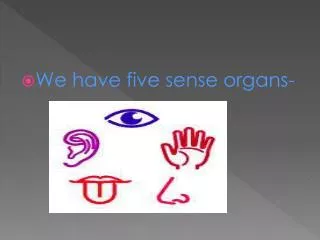
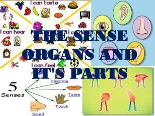
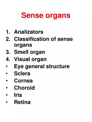
Special Senses Taste Smell Hearing Balance Vision
Physiology of Taste • Gustation is the sensation of taste resulting from the action of chemicals on the taste buds • To be tasted, molecules must dissolve in saliva • 5 primary sensations: salty, sweet, sour, bitter & umami (taste of amino acids) • all tastes can be detected throughout the tongue surface • Neurological Pathway • Taste bud to VII or IX or X to PARIETAL lobe (where in the parietal lobe?)
The Chemical Sense -- Taste • Taste is actually smell • Sweet Salty Sour Bitter • (Sweet Sal, so bitter)
The Chemical Sense -- Smell • Cranial nerve 1 (Olfactory) • Direct outgrowth of brain matter • smell is highly sensitive (more so in women) • distinguish as many as 10,000 odors • CN 1 goes through the cribriform plate on the __________ bone.
The Olfactory • Neurological Pathway of CN 1 • CN1 to Olfactory cortex of TEMPORAL lobe • No fibers go through the thalamus • ANOSMIA- (ann-os-me-a) loss of smell and also taste • CN 1 neurons are replaced every month • Something smells fishy about that, can you sniff it out?
Outer Ear • Fleshy auricle (pinna) directing air vibrations down auditory canal (external auditory meatus) • The ear is housed in the temporal bone
Ear Anatomy • External ear • TYMPANIC MEMBRANE • Middle ear • Auditory ossicles= Malleus, Incus, Stapes (MIS) • Eustachian tube (Auditory tube) goes from middle ear to nasopharynx • opens during swallowing or yawning to equalize air pressure on both sides of eardrum • Inner ear • Cochlea • Semicircular canals
Physiology of Hearing -- Middle Ear • Events: • Tympanic membrane vibrates ossicles (M-I-S) • Stapes moves oval window • Waves in perilymph of Cochlea result • Waves go from Scala Vestibuli around Helicotrema to Scala Tympani • Movement of perilymph stimulates ENDOLYMPH which stimulates VIII
Equilibrium • Control of coordination and balance • Static equilibrium is the perception of a static body moving • perceived by saccule & utricle • Dynamic equilibrium is perception of where you are in space • perceived by semicircular ducts
The Saccule and Utricle • Saccule & utricle • Patch of hair cells buried in a blue Jell-O (otolithic membrane) • This is weighted with rocks on top called otoliths Otoliths Blue Jell-O
Saccule and Utricle • With the head erect, stimulation is minimal, but when the head is tilted, weight of membrane bends hairs (static equilibrium) • When car begins to move at green light, linear acceleration is detected since heavy otolith lags behind (one type of dynamic equilibrium)
Physiology of Balance • Equilibrium= two types • STATIC, DYNAMIC • STATIC= Saccule and Utricle • Otolithic organ, rocks in your head • Rocks tilt with head movement • DYNAMIC= Semicircular canals • PITCH, ROLL, YAW • Semicircular canals hold fluid that stimulates VIII at AMPULLAS
CLINICAL – Ear Pathology • DEAFNESS • Conduction= MIS ossification, noise • Nerve= disruption of nerve • MENIERE’S SYNDROME • Increased endolymph, tinnitus, vertigo, deafness, debilitating • Cranial treatment= endolymph is similar to CSF • TINNITUS= put a ring in your ear
Tear Flow • Tear flow from the lacrimal gland that is located ____ and ____ to the eye • Tears flow into the nasolacrimal duct • Tears contain bactericidal enzyme
The Optical Components • Sclera = white of the eyes • Cornea is transparent avascular covering the iris • Iris = colored part, regulates light with a hole called the Pupil in the middle • Vitreous humor is jelly filling the space between the lens and retina • Aqueous humor is clear serous fluid filling area in front of lens (between lens and cornea) (Aqueous is Anterior) • Lens focuses image • accommodation of lens • Near vision: contraction of ciliary muscle relaxes suspensory ligaments which allows lens to thicken. • light is refracted more strongly & focused onto retina • Describe the physiology of far and near vision.
Anatomy of Internal Eye • Retina forms as an outgrowth of the brain • Red eyes on photos • Optic disc= site where CN II exits, blind spot, blood vessel also exit here • Rods= Black and white and night vision • Cones= color (Baskin Robbins on a cone) • Macula= exact center of vision • Visual purple = visual pigment of the rod cells
Test for Blind Spot • Optic disk or blind spot is where optic nerve exits the posterior surface of the eyeball • no receptor cells are found in optic disk • Blind spot can be seen using the above illustration • in the right position, stare at X and red dot disappears • Visual filling is the brain filling in the bar across the blind spot area (implication for life perceptions)
How Glasses Work • Hyperopia is farsighted (eyeball too short) • correct with convex lenses • Myopia is nearsighted (eyeball too long) • correct with concave lenses • “I’m near to my heart.”
VISUAL PATHWAY • Retina to chiasm through thalamus to Occipital lobe • Pathway divided into three parts: • OPTIC NERVE= Eye to chiasm • OPTIC TRACT= Chiasm to Thalamus • OPTIC RADIATION= Thalamus to Occipital lobe
Visual Pathway LesionsBitemporal Hemianopia • Cause = Pituitary Tumor. Famously afflicted was Goliath, the giant, of the bible.
Visual Pathway LesionsHomonomous Hemianopia • A cause = tumor of the Thalamus
CLINICAL – Eye Pathology • Macula Degeneration • Glaucoma • Cataracts • Presbyopia • Color blindness