Download
1 / 50
510 likes | 576 Vues
Explore the fascinating world of the visual system, from the nature of light as electromagnetic energy to the intricate structure of the human eye. Learn how light energy is transformed into neural responses, the functions of different eye parts like cornea, iris, and lens, and the role of rods and cones in vision.

E N D
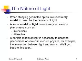
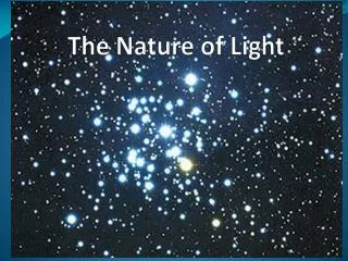
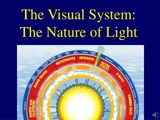
Electromagnetic Energy • An energy spectrum that includes X-rays, radar, and radio waves • A small portion of the spectrum includes light visible to the human eye • Light can be described as both a particle and a wave
Hue • Hue is the color of light as determined by the wavelength of the light energy • Includes: red, orange, yellow, green, blue, indigo and violet (ROY G. BIV) • The eye can detect 7 million separate hues
Intensity/Amplitude • The brightness of light as determined by height of the wave • The taller the wave, the brighter the color
Purpose of the visual system • Transduction: transform light energy into an electro-chemical neural response (action potential & synaptic transmission) • represent characteristics of objects in our environment such as size, color, shape, and location
Cornea • The clear bulge on the front of the eyeball • Begins to focus the light by bending it toward a central focal point • Protects the eye
Iris • A ring of muscle tissue that forms the colored portion of the eye; creates a hole in the center of the iris (pupil) • Regulates the size of the pupil by changing its size--allowing more or less light to enter the eye
Pupil • The adjustable opening in the center of the eye that controls the amount of light entering the eye (surrounded by the iris) • In bright conditions the iris expands, making the pupil smaller. • In dark conditions the iris contracts, making the pupil larger.
Lens • A transparent structure behind the pupil; focuses the image on the back of the eye (retina) • Muscles that change the thickness of the lens change how the light is bent thereby focusing the image (Accommodation) • Glasses or contacts correct problems in the lens’ ability to focus.
The Cosmic Flower Does this picture seem to pulsate? Because the lens of your eye is not perfectly round some parts of what you look at are blurry. Your eyes make micro movements to try to put this entire picture into focus and this creates the pulsation.
The lens correctly focuses the image onto the back of the eye (retina).
(Myopia) The lens correctly focuses the image onto the back of the eye (retina). Misshapen eye causes lens to focus light rays from a distant object in front of the retina. Can see near but not far.
The lens correctly focuses the image onto the back of the eye (retina). (Myopia) Misshapen eye causes lens to focus light rays from a distant object in front of the retina. Can see near but not far. Misshapen eye causes lens to focus light rays from near objects past the retina. Can see far but not near. (Hyperopia)
Other Causes of Poor Vision Astigmatism – Uneven curvature of the cornea causes multiple focus points/images on the retina resulting in blurry vision.
Other Causes of Poor Vision • Presbyopia – form of farsightedness caused when lens becomes brittle & inflexible. Usually starts to happen in your early 40’s Reading glasses will correct this.
Farsightedness Nearsightedness
Retina • Light-sensitive surface with cells that convert light energy to nerve impulses • At the back of the eyeball • Made up of three layers of cells • Receptor cells (Rods & Cones) • Bipolar cells • Ganglion cells
Fovea • The central focal point of the retina • The spot where vision is best (most detailed – visual acuity) • Only cones are found in the Fovea
Receptor Cells(Rods & Cones) • These cells are present in every sensory system to change (transduce) some other form of energy into neural impulses. • In sight they change light into neural impulses the brain can understand. • Visual system has two types of receptor cells – rods and cones
Rods • Visual receptor cells located in the retina • Can only detect black and white • Respond to less light than do cones • Located around the fovea. (remember pen-top demo from class)
Cones • Visual receptor cells located in the retina • Can detect sharp images and color • Need more light than the rods • Many cones are clustered in the fovea at the center of the retina. (remember pen-top demo from class)
Rods Cones Watch Blue Man Group’s Rods & Cones Performance
Distribution of Rods and Cones • Cones—concentrated in centerof eye (fovea) • approx. 6 million • Rods—concentrated in periphery • approx. 120 million • Stare at a word and you’ll notice the others around it become blurred. • (Clear word seen with cones, blurry area seen with Rods)
The Hermann Grid Are there gray dots between the squares? Rods in the periphery are responsible for this. When you look at an area directly there is no dot because you are using your cones but the periphery has dots because the rods are trying to do two things, show you there is a dark area and a light area.
Optic Nerve/Blind Spot/Optic Disc • The nerve that carries visual information from the eye to the thalamus then on to the occipital lobes of the brain • Blind Spot has NO rods or cones
Blind Spot • The point at which the optic nerve travels through the retina to exit the eye (Optic Disk) • There are no rods and cones at this point, so there is a small blind spot in vision. (do demo on page 174) • We don’t notice our blind spot because each eye compensates for the other or your brain “fills in” the missing background info. (Top-down process & Gestalt Theory) • Want more Blind Spot demos? Click HERE Cover your right eye and stare at the can as you move closer to the screen. Notice the spider disappear in your peripheral vision?
Processing Visual Information • Rods & Cones transform light into action potential/synaptic transmission. • Bipolar cells—neurons that connect rods and cones to the ganglion cells • Ganglion cells—neurons that connect to the bipolar cells, their axons form the optic nerve • Optic chiasm—point in the brain where the optic nerves from each eye meet and partly crossover to opposite sides of the brain
Bipolar Cells • Cells that form the middle layer in the retina • Gather information from the rods and cones and pass it on to the ganglion cells • Hundreds of Rods feed into 1 Bipolar Cell • 1 to 2 Cones feed into 1 Bipolar Cell. • This is why Cones have better visual acuity/clarity but Rods collectively can see better in dim lighting
Ganglion Cells • Pass the information from the bipolar cells through their axons • Together these cells form the optic nerve. • The top layer of the cells in the retina
Visual PathwayFrom the eye to the brain Light travels through… Cornea – Pupil – Lens – Fovea (retina) – Rods/Cones – Bipolar Cells – Ganglion cells (movement & light /color & detail) – Optic Nerve (blind spot) – Optic Chiasm (crossover point) – Thalamus – Occipital Lobe (Primary Visual Cortex)
How Can I Possibly Remember All of That in Order? Cherry = Cornea Pie = Pupil Loses = Lens Flavor = Fovea (Rods & Cones) Because = Bipolar Cells Grandma = Ganglion Cells Never = Optic Nerve Cleans = Optic Chiasm The = Thalamus Oven = Occipital Lobe Don’t like this one? Create Your Own!!!
Primary Visual Pathway:Thalamus processes info about form, color, brightness & depth (What Pathway) Secondary Visual Pathway:Midbrain processes info about the location of an object (Where Pathway) – feature detectors respond to things like angles, edges, lines & movement
Parallel Processing of Vision • Brain simultaneously processes information about Motion, Form, Depth & Color then compares it to memories you have stored.
Visual Adaptation (a) A projector mounted on a contact lens makes the projected image move with the eye. (b) Initially the person sees the stabilized image, but soon she sees fragments fading and reappearing.
Visual Impairment • Play “Smart Glasses” (8:13) Segment #9 from Scientific American Frontiers: Video Collection for Introductory Psychology (2nd edition). Watch in Class