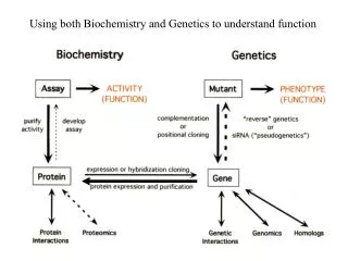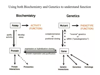
Using both Biochemistry and Genetics to understand function
320 likes | 375 Vues
Explore the synergy of Biochemistry and Genetics in deciphering the intricate mechanisms of DNA Replication, including enzyme functions, fidelity, regulation, and structure. Delve into the challenges and breakthroughs in studying semiconservative duplication speed and fidelity.

Using both Biochemistry and Genetics to understand function
E N D
Presentation Transcript
DNA Replication: The Task and Challenge Semiconservative Duplication Speed: very rapid duplication of every nucleotide (ex: 6 x 10⁹ bp in 8 hrs in humans) Fidelity: extremely low error rate (~1/109 nucleotide error rate) Count: exactly two copies of every sequence per cell cycle Regulation: coordination with other chromosomal events (eg.mitosis, repair, recombination, transcription, chromatin packaging)
Enzymology of DNA Synthesis: DNA Polymerases dNTP precursor - pyrophosphate release provides energy Instructed by single-stranded template - senses complementarity of new nucleotide Primer requirement* - senses complementarity of primer 5’ > 3’ polymerization off primer* - extension off 3’ hydroxyl - moving 3’> 5’ on template * enhances fidelity by allowing error correction
Assaying DNA Polymerase Activity In principal: Monitor incorporation of radioactive nucleotide precursors ( ) into acid insoluble form (physically separable on filter) In practice: Can be difficult to devise the right assay conditions when you do not know the precise nature of the activity Initial conditions used were really assaying a complex mixture of activities: E. coli extract - source of polymerase activity but also kinase and nuclease activity 3H Thymidine - converted to thymidine triphosphate by kinases in extract DNA - intended as nuclease decoy but nucleases convert to primer-template and source of A,G,C nucleotides First Experiment: 50 out of 1 million cpm insoluble Ten Years Later: purify DNA Polymerase I and figure out enzyme requirements
DNA Polymerase Structure and Catalysis Crystal structure of bacteriophage T7 DNA Polymerase complexed with primer-template and dNTP Two Mg++ ions positioned by conserved acidic residues catalyze reaction Primer Template Rest of enzyme positions primer-template and dNTP and ensures catalysis only occurs with proper “fit” Structure resembles a right hand
DNA Pol I has 3’ > 5’ Exonuclease Activity Careful quantitative analysis of biochemical activity can suggest biological function * T * T T T T T T T 3’ 5’ Exo Assay: DNA Pol I 5’ 3’ AAAAAAAA AAAAAAAA 5’ 5’ 3’ 3’ no dTTP exo activity is slow relative to pol activity exo activity is enhanced by stalling pol activity or making 3’ end single-stranded 3’ mismatch generates both conditions * * T T T T T T T T T T T T Proofread Assay: 3’ 5’ 5’ DNA Pol I 3’ AAAAAAAA AAAAAAAA 5’ 3’ 5’ 3’ + dTTP * T T T T T T T T T T C T 3’ 5’ 5’ DNA Pol I 3’ AAAAAAAA AAAAAAAA 5’ 3’ 5’ 3’ + dTTP * C mismatch specific exo activity under normal pol conditions both pol and exo activities are sensing primer-template pairing
The Polymerase and Exonuclease Activities of Replicative DNA Polymerases Reside in Distinct Domains 2- Mode Model for Polymerase Function Polymerase Active Site ~ 30Å Exonuclease Active Site Polymerizing Editing Movement between P and E sites requires primer-template unwinding translocation of 3’ end
DNA Pol I is not the replicative DNA polymerase in E. coli Illustrates importance of genetics for establishing functional relevance in cell Use biochemical assay to screen for mutants lacking DNA polymerase activity plate mutagenize mutant E. coli E. coli assay dNTP incorporation into DNA extracts from single mutant colonies mutant 3473 (polA1) has <1% wt activity polA1 phenotypes: normal growth;repair deficient Purification of residual polymerase activity from polA1 yields DNA Pol II and Pol III Genetics later establishes that DNA Pol III is the primary replicative polymerase
Purification of DNA Pol III: Different Template, Different Assay, Different Activity Introducing the concept of holoenzymes and modular enzyme subassemblies
Fidelity Overview Contributions to E coli DNA Replication Fidelity Fidelity Comparisons Error Rate Product Size Speed Error rate DNA Replication Intrinsic Fidelity (polym) 10-3 - 10-4 500 bp/sec 5 x 10⁶ 10-9 - 10-10 (Prokaryotes) (E. coli) (sensing dNTP complementarity to template) 6 x 10⁹ 50 bp/sec (humans) (Eukaryotes) Exonuclease Proofreading (polym) 10-2 - 10-3 (sensing primer complementarity to template) 1 x 10¹¹ (lily) Mismatch Repair (post polym) 10-2 RNA Transcription 30 bp/sec 10-4 103 - 106 (sensing complementarity of two strands) (distinguishing parental and daughter strands) Protein Translation 20 aa/sec 10-4 102 - 103 Overall Replication Fidelity 10-8 - 10-9
Models for Polymerase Discrimination H-bonding (binding energetics) Outside the active site, unpaired nucleotides are H-bonded to H2O. Inside the active site these H-bonds can be replaced by WC base pairing but only incompletely replaced by mismatch pairing Mismatch H bonding can also exacerbate steric and stacking clashes (see below) Steric Constraints (structure/geometry) Imposed by enzyme’s “induced fit”, which can test for precise base pair geometry, proper base stacking, and correct primer template fit. How to Distinguish Mismatch versus Correct Base Pair Geometry From Crystal Structure WC bp mismatch WC bp mismatch mismatch Global structure of helix is not greatly perturbed But there are: differences in C1’ - C1’ distance and C1’ bond angles protrusions of bases into major groove loss of universal H acceptor positions in minor groove
C C C k k k pol conf conf C C K K C dNTP D E DNA D N C E DNA PP N+1 i * C dNTP E DNA N 1 3 2 E DNA N I I I k k k pol conf conf I K D I K I PP E DNA D N+1 i * I dNTP E DNA N I dNTP E DNA N Intrinsic Fidelity: Potential Base Pair Discrimination for dNTP at Three Stages Of the DNA Polymerization Reaction Cycle Arrow thickness roughly corresponds to rate constant Reaction pathway for correct nucleotide Reaction pathway for incorrect nucleotide Rapid dNTP Binding Pseudo-equilibrium Slow Conformational Change “Induced Fit” Polymerization Reaction Example: T7 DNA Polymerase (Other polymerases discriminate differently at each stage) -1 > 8mM 300 s ~ ~ ~ ~ 1500x 400x Rapid and Not Measured -1 20 µM 0.2 s
C C k k pol conf C E DNA PP C C K K N+2 i C dNTP E DNA D D N * C dNTP E DNA N 1 3 2 I C I C I k k conf pol C E DNA N+1 I * C C E dNTP dNTP DNA I I E DNA E DNA N+1 N+1 N+1 Error Correction: Primer requirement allows kinetic discrimination sensitive to base pairing of recently incorporated nucleotides Arrow thickness roughly corresponds to rate constant Fast reaction pathways for correct primer with correct nucleotide Slow reaction pathways for incorrect primer with correct nucleotide I E DNA PP N+2 i Rapid dNTP Binding Pseudo-equilibrium Slow Conformational Change “Induced Fit” Polymerization Reaction
C C E E DNA DNA C C k k k N N exo pol conf C E DNA PP C C K K N+2 i C dNTP E DNA D D N * C dNTP E DNA N 1 3 2 I C I C I k k conf pol I k exo C E DNA N+1 I E DNA PP I * C C N+2 i E dNTP dNTP DNA I I E DNA E DNA N+1 N+1 N+1 Error Correction: Exonuclease activity allows the polymerase’s kinetic discrimination to lead to different primer fates Arrow thickness roughly corresponds to rate constant Fast reaction pathways for correct primer with correct nucleotide Slow reaction pathways for incorrect primer with correct nucleotide
C C E E DNA DNA N N C E DNA PP N+2 i C E DNA N+1 I E DNA PP N+2 i I E DNA N+1 Error Correction: Kinetic manipulation of molecular choice based on complementarity of primer Arrow thickness roughly corresponds to rate constant When a correct nucleotide is incorporated, 3’>5’ exonuclease activity is much slower than 5’>3’ polymerase activity. Addition of the next nucleotide is kinetically favored. exo pol pol When an incorrect nucleotide is incorporated, disruption of the primer greatly slows 5’>3’ polymerase activity for the next nucleotide (and slightly increases 3’>5’ exonuclease activity). Excision of the incorrect nucleotide is kinetically favored. exo
5’-TAGCTTC 5’-TAGCTTCG 5’-TAGCTTCGA 5’-TAGCTTC 5’-TAGCTTC 5’-TAGCTTCA 3’-ATCGAAGCTCATG 3’-ATCGAAGCTCATG 3’-ATCGAAGCTCATG 3’-ATCGAAGCTCATG 3’-ATCGAAGCTCATG 3’-ATCGAAGCTCATG A A Error Correction: Kinetic manipulation of molecular choice based on complementarity of primer Black arrow thickness roughly corresponds to relative rate constant Light blue arrow thickness roughly corresponds to relative flux exo pol pol exo
One General Strategy for Fidelity: Kinetic manipulation of molecular choice between irreversible forward and discard pathways The choice is ultimately determined by the relative flux of molecules that proceed down the two competing pathways (light blue arrow) Elimination Pathway irreversibility usually requires some chemical energy expenditure (e.g dNTP hydrolysis), which could be coupled to either pathway or to a reaction step preceding these pathways discard forward Cognate Substrate Correct Product forward Noncognate Substrate Incorrect Product For each substrate, the molecular flux (and hence molecular choice) is determined by the ratio of the forward to discard rate constants (black arrows) for that substrate. For cognate substrates this ratio should favor the forward reaction. For noncognate substrates, the ratio should “flip” to favor the discard pathway. discard Elimination In principal, just one or both pathways could discriminate between cognate and noncognate substrates, i.e. change rate constants with substrate. In practice, nature often discriminates with both.
How DNA Polymerases Check for Proper Base Pairing Geometry Crystal Structure Evidence for “Induced Fit” DNA Polymerase contacts minor groove of primer-template Stacking Interaction Primer Template Polymerase + Primer-Template Polymerase + Primer-Template + dNTP Base pair fit is “tested” before polymerization Base pair fit is still “tested” after polymerization Only W-C base pairs allow proper stacking Purple: Interaction surface with DNA polymerase Induced fit positions nucleotide, primer 3’, metal ions Green: Universal H-bond acceptors H-bonding with DNA polymerase
Many DNA polymerases in the cell have nonreplicative roles Prokaryotic DNA Polymerases (E. coli) DNA Replication(RNA primer removal); DNA repair DNA repair DNA Replication DNA repair; TLS; adaptive mutagenesis TLS (translesion synthesis) Pol I Pol II (Din A) Pol III holoenzyme Pol IV (Din B) Pol V (UmuC, UmuD’2C) Eukaryotic DNA Polymerases DNA Replication (Primer Synthesis) Base excision repair Mitochondrial DNA replication/repair DNA Replication; nucleotide and base excision repair DNA Replication; nucleotide and base excision repair DNA crosslink repair TLS Meiosis-associated DNA repair Somatic hypermutation TLS Error-free TLS past cyclobutane dimers TLS, somatic hypermutation TLS Pol a Pol b Pol g Pol d Pol e Pol q Pol z Pol l Pol m Pol k Pol h Pol i Rev1 Most of the nonreplicative polymerases have low fidelity and are error-prone because they tolerate non-WC bp and lack 3’>5’ exo activity Low fidelity is needed to bypass template lesions that are stalling replication Low fidelity may be used to increase genetic variation in special circumstances
DNA Synthesis Occurs Semi-Discontinuously at Replication Forks Leading daughter strand: polymerase moves continuously in same direction as replication fork Lagging daughter strand: polymerase moves discontinuously in opposite direction as replication fork 5’ Fork Movement C 3’ B lagging A 3’ 5’ leading 5’ 3’ Synthesis with discontinuous lagging strand pieces (okazaki fragments) requires repeated: A. priming B. replacement of primed sequence C. ligation Okazaki fragment length: prokaryotes 1000 - 2000 nt; eukaryotes 100-200 nt
Structural Analysis of In Vivo DNA Replication Intermediates Replication is more complex than just DNA polymerization Replication is localized to forks (Autoradiograph E. coli DNA) New DNA synthesis is small (pulse label and size) Small SS gaps at forks (EM replicating l DNA) daughter alkaline sucrose gradient analysis can distinguish SS from DS DNA small DS DNA (leading) SS DNA (lagging) fork parent SS DNA (lagging) DS DNA (leading) fork daughter Suggests replication initiates from single site but can’t say that all molecules initiate from same site Higher resolution analysis of okazaki fragments show 8-10nt RNA at 5’ end linked 5’ > 3’ to DNA Antiparallel orientation of SS DNA consistent with presumptive leading/lagging strand assignments
Replication Fork Tasks and the Activities That Perform Them Activity Task helicase unwind parental strands primase begin DNA synthesis SSBP stabilize SS DNA polymerase synthesize DNA clamp loader/clamp ensure processivity unlink parental strands topoisomerase nuclease/polymerase replace primer ligase connect okazaki fragments
Structural “Solution” to DNA Replication “. . . each chain acts as a template for the formation on to itself of a new companion chain.” - Watson & Crick 20 Å 34 Å Structural Implications 1. Complementarity semiconservative model 2. Antiparallel strandspotential asymmetry if replication proceeds along helix in one direction 3. Double helix must disentangle interlinked parental strands during semiconservative replication ref 1
Demonstrating 5’3’ Chain Growth polymer growth IF 5’3’ correct + + PPi P-P-P P-P-P P-P-P OH OH OH P P P P P P P 5’ 3’ 5’ 3’ polymer growth IF 3’5’ correct + + PPi P-P-P P-P-P P-P-P OH P P P OH OH P P P P 5’ 3’ 5’ 3’ Test: Use dideoxy nucleotide to ask if 3’ OH required for monomer incorporation? Result: Dideoxy is incorporated but subsequent polymerization is blocked
Polymerization via Head Growth vs Tail Growth Head Growth: activated high energy bond end of polymer drives polymerization Tail Growth: activated high energy bond end of monomer drives polymerization
5’>3’ allows “discard” exo pathway to return to polymerization 5’ > 3’ polymerization (head growth) 3’>5’ Exo 3’ > 5’ polymerization (hypothetical tail growth) 5’>3’ Exo
Using Biochemical Assays to Define Biochemical Functions Assay must distinguish or physically separate products from substrates Polymerization: conversion of radioactive nucleotide from acid soluble to acid precipitable Nuclease: conversion of incorporated radioactive nucleotide from acid precipitable to acid soluble Quantification is important for inferring biological relevance 3’ > 5’ exonuclease negates the polymerization reaction, but is generally much slower DNA Pol I’s poor polymerization raised the possibility that it was not the replicative helicase Small differences in assay conditions can define different activities gapped/nicked template defines Pol III core activity primed single-stranded template defines Pol III holoenzyme activity Complications of assaying crude extracts (beware of wasting clean thoughts on dirty enzymes) can be detecting multiple types of activities and be affected by multiple competing activities can be detecting multiple similar activities
The Awesome Challenges of Genetics polA mutant revisited Lecture 1: polA1 mutant with <1% assayable DNA Pol1 activity have relatively normal replication Cairns concludes DNA Pol1 is not important for DNA replication Lecture 2: DNA Pol1 plays a role in okazaki fragment maturation, an important part of replication What happened to the awesome power of genetics? Caveats applied to polA1 mutants Caveats about gene analysis Limitations in Phenotypic Analysis Pol I activity in living polA1 cells may be greater than that measured in vitro (in extracts) as mutant protein may be more labile or inhibitable in the harsher in vitro setting than in vivo. E.coli has an estimated 300 molecules of DNA Pol I, most used in DNA repair. Fewer molecules are needed for the 2 replication forks, so the residual activity in a polA1 mutant may be sufficient. Note, although polA1 has an early nonsense mutation, read-through of the nonsense codon is suspected of generating the residual Pol I activity Excess Activity/Leaky Allele Redundancy We can eliminate the first two caveats with a null mutant, butthe polA∆ mutant is still viable in minimal media (although not in rich media, where the demands for rapid DNA replication are greater). In this mutant Pol II or Pol III is thought to substitute (poorly but sufficiently) for Pol I in OF maturation With all these caveat, what is the evidence that DNA Pol1 is important for OF maturation and DNA replication? polA12 ts mutant accumulates increased OFs at restrictive temp (similar to the ts lig4 mutant) polA12lig4 double mutant not only accumulates OFs but rapidly ceases DNA synthesis at restrictive temp
General Strategies for Isolating DNA Synthesis Mutants 1) Screen: assay macromolecular synthesis in vivo in ts-lethal mutants soon after shift to restrictive temp • DNA synthesis– (3H Thymine incorp.) • RNA synthesis + • Protein synthesis + • Nucleotide synthesis + • 2) Selective enrichment: ts mutants that fail to incorporate poisonous • nucleotide analog during transient shift to restrictive temp wash out 5-BU 5-BU 32° 42° 42° 32° UV Can recycle survivors to further enrich WT incorporate 5-bromouracil (5-BU) - UV sensitive Replication mutants poorly incorporate 5-BU - survive
Lots of mutants but limited in vivo assays to characterize generation time Log DNA synthesis Time shift to restrictive temperature Slow Stop: involved in initiation ongoing replication continues new rounds blocked Quick Stop: involved in elongation ongoing replication blocked
Mismatch Repair: Correction of Replication Errors E. coli mismatch repair DNA- both parental strands methylated at GATC sites daughter strand transiently unmethylated after replication MutS - recognizes mispaired bp by susceptibility to kinking MutH - recognizes nearby GATC MutL - association with MutS and MutH stimulates MutH to nick unmethylated daughter strand (basis of strand bias) Exonuclease and helicase II, directed by MutS and MutL excise daughter strand from nick to mispaired bp DNA polIII, clamp, clamp-loader, and SSB synthesize replacement DNA MutS bound to mispaired DNA DNA Dam methylase eventually fully methylates GATC sites so both strands are marked as parental for next round of replication