Download

1 / 124
1.28k likes | 1.66k Vues
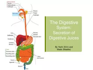
Organs of the Digestive System. For student copy. The Mouth. aka oral or buccal cavity cheeks form lateral walls internally covered by mucous membrane: nonkeratinized , stratified sq epith wall of cheeks: buccinator muscle then subq & skin lips or labia surround opening

E N D
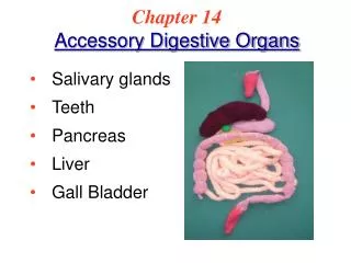
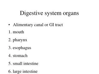
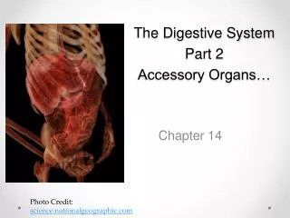
Organs of the Digestive System For student copy
The Mouth • aka oral or buccal cavity • cheeks form lateral walls • internally covered by mucous membrane: nonkeratinized, stratified sq epith • wall of cheeks: buccinator muscle then subq & skin • lips or labia surround opening • inner surface of each lip attached to its gum by a midline fold of mucous membrane called a labial frenulum “small bridle”
Mouth - 2 • vestibule: space between buccal mucosa & teeth • oral cavity proper: space that extends from gums & teeth fauces: opening between oral cavity & pharynx • hard palate: anterior portion of roof of mouth • maxillae & palatine bones form bony partition between oral & nasal cavities • covered by mucous membrane
MOUTH - 3 • soft palate: forms posterior portion of roof of mouth • muscular partition between oropharynx & nasopharynx • uvula : hangs from free border of soft palate • when swallowing soft palate & uvula drawn superiorly preventing food & liquids from entering nasal cavity
Salivary Glands • release saliva into oral cavity • 4 sets: • Parotid glands (“near ear”) • between masseter & skin • parotid duct secretes saliva into vestibule opposite 2nd molar • Submandibular glands • floor of mouth/ enter just lateral to lingual frenulum • Sublingual glands • under tongue • lesser Sublingual glands: floor of mouth
Saliva • 99.5% water • 0.5% solutes: • ions • urea & uric acid • mucus • Ig A • lysozyme (bacteriostatic enzyme) • salivary amylase: digestive enzyme acts on starch
Salivation • controlled by ANS • average adult secretes 1000 – 1500 mL/day • parasympathetic stimulation promotes continuous secretion keeps mouth moist & lubricates tongue & lips during speech • saliva is then swallowed moistening esophagus most water is reabsorbed • sympathetic stimulation dominates if stressed mouth dry • dehydration: secretion stops to conserve water
Mumps • inflammation & enlargement of parotid glands • pain, malaise, fever • swelling on affected side
Tongue • skeletal muscles covered by mucous membrane forming floor of oral cavity • median septum separates tongue into symmetric ½ s (attaches to hyoid bone) • lingulumfrenulum • limits movement posteriorly • if abnl short: “tongue-tied” • each ½ composed of extrinsic & intrinsic muscles • extrinsic: origins out of tongue/ insert to CT in tongue: move tongue side-to-side/ anchor tongue • intrinsic: origin & insertion in tongue: alter shape & size of tongue for speech & swallowing
Tongue - 2 • dorsum (upper surface) & lateral surfaces covered with papillae • projections of lamina propria covered with keratinized epithelium • some contain taste buds • others touch receptors • all increase friction between tongue/food • lingual glands secrete mucous & a watery serous fluid that contains enzyme lingual lipase: acts on triglycerides
Taste Buds • most on tongue, few on soft palate, pharynx, & epiglottis • each taste bud has 3 types epith cells: • supporting cells • surround ~50 receptor cells • gustatory receptor cells • single microvillus • basal cells
Taste Buds - 2 • each taste bud has 3 types epith cells: • supporting cells • surround ~50 receptor cells • gustatory receptor cells • single microvillus from each = gustatory hair extends thru a taste pore (opening in taste bud) • basal cells • stem cells @ edge of taste bud • produce supporting cells that then develop into gustatory cells (each lasts ~10 days)
Teeth • dentes • in alveolar processes of mandible & maxillae • covered by gingivae: “gums” • lined by peridontal ligament: anchors tooth to socket • parts of a tooth: • Crown • Root • Neck
Crown of Tooth • visible portion, above level of gums • interior made of dentin: calcified CT • gives shape & rigidity to tooth • harder than bone • covered by enamel • Ca++ phosphate & carbonate • hardest substance in body • protects tooth from: • wear & tear of chewing • acids that could dissolve dentin
Pulp Cavity • w/in dentin • pulp: CT with blood, lymph & nerve supply to tooth • extension thru roots = root canals opening @ base for vessels/nerve to enter/exit tooth
Root of Tooth • below gums • covered by cementum
2 Dentitions • Deciduous teeth • baby or primary teeth • ~6 mos 24/mo 20 total • lost age 6 - 12 • Secondary teeth • 32 • begin to erupt ~ 6 adult
CHEMICAL Digestion in the Mouth MECHANICAL mastication: chewing food manipulated by tongue/ground by teeth & mixed with saliva bite of food reduced to soft, flexible, easily swallowed mass = bolus Salivary amylase initiates breakdown of starch into di- & trisaccharides, shorter polymers only monosaccharides can be absorbed 2. Lingual lipase becomes activated in acid pH of stomach
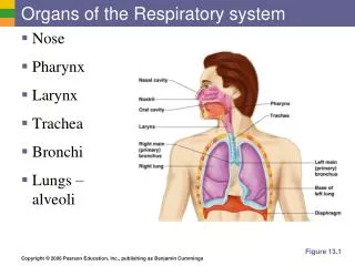
Pharynx • funnel-shaped tube extends from internal nares esophagus posteriorly & to the larynx anteriorly • skeletal muscle covered by mucous membranes • 3 parts: • Nasopharynx: functions only in respiration • Oropharynx: digestive + respiratory functions • Laryngopharynx: digestive & respiratory
Esophagus • collapsable muscular tube • posterior to trachea • begins @ inferior end of laryngopharynx passes thru mediastinum pierces diaphragm (opening called esophageal hiatus) ends in superior portion of stomach
Histology of the Esophagus • mucosa: • nonkeratinized stratified sq epith • lamina propria • Muscularismucosae (smooth muscle) • submucosa: areolar CT • muscularis: • upper 1/3 skeletal • mid 1/3 skeletal & smooth • lower 1/3 smooth
Ends of Esophagus • muscularis thickens forming: • upper esophageal sphincter (UES) • skeletal • regulates movement of food from pharynx esophagus • lower esophageal sphincter (LES) • smooth • regulates movement of food from esophagus stomach
Physiology of the Esophagus • secretes mucus & transports food stomach • No enzymes produced or secreted • No absorption
Deglutition • swallowing • facilitated by secretion of saliva & mucus • involves mouth, pharynx, esophagus • Voluntary stage • bolus of food from oral cavity to oropharynx • stimulates receptors in oropharynx deglutition center in medulla & lower pons effector fibers cause soft palate & uvula to move up to close off nasopharynx AND epiglottis closes off opening of larynx
Deglutition Involuntary Stage 2. Esophageal stage • bolus enters esophagus • peristalsis: progression of coordinated contractions & relaxations of circular & longitudinal layers of muscularis, pushes bolus onward
Stomach • J-shaped enlargement of GI tract • just inferior to diaphragm • connects esophagus duodenum • most distensible part of GI tract • serves as a • mixing chamber • holding reservoir
Stomach Adaptations for Digestion • rugae • mucus glands: • secretion of H+ & Cl- • pepsin • gastric lipase • intrinsic factor • 3-layered muscularis
Histology of the Stomach • 4 basic layers in stomach wall: • (stomach wall is impermeable to most substances) • surface mucosa = simple columnar epith that extend down into lamina propria where they form columns of secretory cells called gastric glands, channels between columns called gastric pits
Mechanical Digestion in Stomach • few minutes after food bolus enters stomach: gentle, rippling, peristaltic movements called mixing waves pass thru stomach q15 – 25 s • macerate food • mix with mucus secretions • results: chyme soupy liquid pylorus
Pyloric Sphincter • slightly open • when chyme down to lower pylorus, each mixing wave forces ~ 3 mLchyme into duodenum = gastric emptying
Chemical Digestion in the Stomach • salivary amylase: • continues to function while food in fundus • when churning forces bolus further into stomach the acid pH inactivates it • lingual lipase: • acid pH activates • triglycerides fatty acids & diglycerides
Chemical Digestion - 2 • H+ & Cl- ions secreted separately by parietal cells • secretion stimulated by: • parasympathetic neurons • gastrin (from G cells) • histamine (from mast cells in lamina propria): receptors on parietal cells = H2 receptors
Stomach Acid • kills many microbes in food • partially denatures proteins • stimulates secretion of hormones that promote flow of bile & pancreatic juice
Pepsin • secreted by chief cells • greatest activity in low pH / inactivates in higher pH of small intestine • secreted as pepsinogen: inactive form of pepsin (so will not break down proteins in chief cells • severs peptide bonds breaking protein smaller peptide fragments • stomach wall protected from pepsin by alkaline mucus secreted by surface cells
Gastric Lipase • splits short-chain triglycerides fatty acids & monoglycerides • most effective @ pH 5-6 (limited role in stomach)
Absorption in Stomach • very little in stomach (epithelial cells impermeable to most substances) • mucous cells do absorb some: • water • ions • short-chain fatty acids • aspirin • alcohol
Stomach • 2 – 4 hrs for food to exit • meal mostly carbs: shortest time • protein – rich meal longer • fat-laden meal longest