Download
1 / 19
560 likes | 2.66k Vues
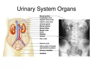
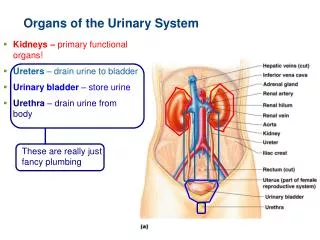
Organs of the Urinary System. Kidneys – primary functional organs! Ureters – drain urine to bladder Urinary bladder – store urine Urethra – drain urine from body. These are really just fancy plumbing. Kidney Anatomy. Retroperitoneal (behind peritoneum), superior lumbar region

E N D
Organs of the Urinary System Kidneys – primary functional organs! Ureters – drain urine to bladder Urinary bladder – store urine Urethra – drain urine from body These are really just fancy plumbing
Kidney Anatomy Retroperitoneal (behind peritoneum), superior lumbar region Right kidney is lower than left Renal hilum leads to renal sinus (medial cavity) Ureters, vessels, and nerves enter/exit at hilum Renal hilum Anterior Peritoneal cavity (organs removed) Peritoneum 3 Supportive tissue layers Renal vein • Renal fascia anterior posterior Renal artery • Perirenal fat capsule Body of vertebra L2 • Fibrous capsule
Regions of the Kidney • Renal cortex—outer region • Renal medulla—inside cortex • Medullary pyramids and renal columns Figure 15.2b
Internal Kidney Anatomy Papilla = tip of pyramid; drains urine into minor calyx Minor Calyces drain urine to major calyces Renal pelvis receives urine from calyces, empties into ureters Renal cortex Renal medulla Major calyx Papilla of pyramid Renal pelvis Minor calyx Renal pyramid in renal medulla Renal column Ureter Fibrous capsule (b) Diagrammatic view Figure 25.3
Kidneys filter blood 1/4th of blood supply passes through kidneys per minute! Renal artery = arterial blood supply (to be filtered) Renal vein = filtered blood drainage Filtration occurs across capillaries = glomerulus in between . . . Be sure to learn these today…
Cortical radiate vein Figure 25.4 Blood vessels of the kidney. Aorta Inferior vena cava Cortical radiate artery Arcuate vein Renal artery Renal vein Arcuate artery Interlobar vein Segmental artery Interlobar vein Interlobar artery Segmental arteries Interlobar artery Arcuate vein Renal vein Cortical radiate vein Renal artery Arcuate artery Renal pelvis Peritubular capillaries and vasa recta Cortical radiate artery Ureter Afferent arteriole Efferent arteriole Renal medulla Glomerulus (capillaries) Renal cortex Nephron-associated blood vessels (see Figure 25.7) (a) Frontal section illustrating major blood vessels (b) Path of blood flow through renal blood vessels
Nephrons = functional units of kidneys ~1 million per kidney Two main parts Glomerulus: a tuft of fenestrated capillaries Renal tubule: Glomerular (Bowman’s) capsule Proximal Convoluted Tubule (PCT) Loop of Henle Distal Convoluted Tubule (DCT) Glomerulus + Glomerular Capsule = Renal Corpuscle
Nephron epithelia Hug glomerulus capillaries Glomerular capsule: parietal layer Renal cortex Basement membrane Renal medulla Renal corpuscle Podocyte • Glomerular capsule Renal pelvis Fenestrated endothelium of the glomerulus • Glomerulus Distal convoluted tubule Ureter Glomerular capsule: visceral layer Kidney Microvilli Mitochondria Proximal convoluted tubule Highly infolded plasma membrane Cortex Proximal convoluted tubule cells Medulla Thick segment Distal convoluted tubule cells Thin segment Loop of Henle • Descending limb • Ascending limb Collecting duct Loop of Henle (thin-segment) cells Principal cell Intercalated cell Thick segment of loop = cuboidal/columnar Collecting duct cells Figure 25.5
Filtration slits Figure 25.9b The filtration membrane. Podocyte cell body Foot Processes (a.k.a pedicels) (b) Filtration slits between the podocyte foot processes
2 types of nephrons… Cortical nephrons – found in cortex, most common (85%) Juxtamedullary nephrons – penetrate medulla via extra long Loop of Henle Juxtamedullary nephrons especially equipped to reabsorb H2O, i.e. concentrate urine
Juxtaglomerular Apparatus (JGA) Regulates filtration and b. p. Granular cells (juxtaglomerular, or JG cells) Enlarged, smooth muscle cells of arteriole(s) Granules contain renin Sense blood pressure (baroreceptors) Macula densa Tall, closely packed cells of ascending limb Chemoreceptors sense NaCl in filtrate Extraglomerular mesangial cells May pass signals b/n macula densa/granular cells • Macula densa cells of the ascending limb of loop of Henle • Extraglomerular mesangial cells • Granular cells Juxtaglomerular apparatus
Renal corpuscle Figure 25.6a Renal cortical tissue and renal tubules. • Glomerular capsular space • Squamous epithelium of parietal layer of glomerular capsule • Glomerulus Artery Distal convoluted tubules (clear lumens) Proximal convoluted tubules(fuzzy lumens due to long microvilli) (a) Photomicrograph of renal cortical tissue (200x)
Figure 25.21a Structure of the urinary bladder and urethra. Peritoneum Ureter Rugae Detrusor muscle Adventitia Ureteric orifices Trigone of bladder Bladder neck Internal urethral sphincter Prostate Prostatic urethra Urogenital diaphragm External urethral sphincter Membranous urethra Spongy urethra Erectile tissue of penis External urethral orifice (a) Male. The long male urethra has three regions: prostatic, membranous and spongy.
Figure 25.21b Structure of the urinary bladder and urethra. Peritoneum Ureter Rugae Detrusor muscle Ureteric orifices Bladder neck Internal urethral sphincter Trigone External urethral sphincter Urogenital diaphragm Urethra External urethral orifice (b) Female.