The Excretory System and Kidneys: Functions & Structure
150 likes | 182 Vues
Learn how the kidneys filter blood, remove wastes, and maintain chemical balance. Explore the urinary system's anatomy, nephron structure, and processes like filtration, reabsorption, and secretion.

The Excretory System and Kidneys: Functions & Structure
E N D
Presentation Transcript
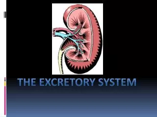
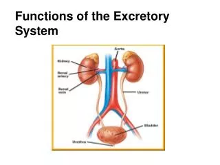
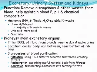
Excretory/Urinary System and Kidneys • Function: Remove nitrogenous & other wastes from blood, help maintain blood P, pH & chemical composition • Ammonia (NH3)- Toxic H2O-soluble N-waste • Urea- H2O-based • Majority of N waste in humans • Uric acid- more solid • Creatinine • Kidneys= main excretory organs • Filter 200L of fluid from bloodstream a day & make urine • Location: dorsal body wall between, near bottom of rib cage • 3 processes of blood purification: • Filtration- using P & a filter to separate substances, creating filtrate • Reabsorption- absorbing useful material back from filtrate • Secretion- transporting substances into forming filtrate
… From the Kidneys…. • Ureters- tube from each kidney to bladder • Continuous with “renal pelvis” • Muscular layer of walls, active roll in transporting urine • Urinary bladder- smooth, collapsible, muscular bag that stores urine • On pelvic floor, just behind pubic symphysis • Transitional epithelium • Urethra- thin-walled muscular tube from bladder to external opening • Internal urethral sphincter- at bladder urethra junction, involuntary, closed when urine not passing through • External urethral sphincter- surrounds urethra as passes through urogenital diaphragm, voluntary skeletal muscle
Micturition (Voiding) • Stretch receptors in bladder wall activate, send signals to spinal cord, 2 Reflexes: • Low stretch- sympathetic nervous system inhibits bladder muscle to keep internal sphincter closed • More stretch- stimulate contraction of external sphincter • When ~200mL of urine has accumulated, afferent motor fibers make contraction of bladder more frequent & urgent • If allowed, voiding reflex initiated • If not voided, contractions subside after about 1 min, once ~200mL more urine accumulates, process repeats • When V exceeds 500-600mL, urge to urinate becomes irresistible
Structure of the Kidney • Bean-shaped, hilum of medial surface for entrance/exit ureter, blood & lymph vessels, & nerves • Internal Anatomy: • Renal Cortex- superficial • Renal Medulla- darker, with multiple cone shaped renal pyramids • Each with many “collecting ducts” leading to a papillae • Renal Pelvis- Collects urine from papillae of renal pyramids, funnels into ureter • Common area for formation of kidney stones • Blood and Nerve Supply: • Large Renal Artery & Vein to each kidney • 1/4 total cardic output directly to kidneys each minute • Artery branches many times, until over 1mil vessels going to each “nephron” in each kidney • Renal Plexus- autonomic nerves supplying kidneys & ureters, regulating vasomotor fibers & blood flow to & urine forming role of kidney
Nephrons • Structural & functional unit of kidney • Each kidney has >1mil, each with own arteriole, each connected to 1 of 1000s of collecting ducts, leading from medulla to pelvis • Anatomy: • Glomerulus: Clump of capillaries • Delivers blood for filtration, artery feeding glomerulus also wraps around tubule to facilitate processing of urine • Renal tubule: Collects & processes (reabsorption & secretion) filtrate • Bowman’s Capsule- cup-like end, surrounds glomerulus • Proximal Convoluted Tubule- PCT- (coils as leaves Bowman’s) • Loop of Henle (descending and ascending limbs) • Distal Convoluted Tubule- DCT- (coils after loop, continues to connect to a collecting duct)
Filtration • Hydrostatic P drives fluids & some solutes out of blood in glomerulus, into Bowman’s capsule • Passive • Filtrate includes H2O, NaCl, NH3, urea, uric acid, glucose, amino acids, etc • Kidneys make approx 180L filtrate a day, less than 1% actually leaves body • Membrane of capillaries in glomerulus 1000x more permeable than others • Walls of Bowman’s capsule also very permeable • Blood P in glomerulus much higher than in other capillaries
Regulation of Filtration • Intrinsic- smooth muscles of arterioles contract/relax, controlling flow into glomeruli, give tubules enough time to make adjustments in filtrate • Extrinsic- Sympathetic nervous system adjusts blood vessel diameter in response to blood P or “fight-or-flight” response. Hormones adjust blood P by stimulating reabsorption of Na+, increasing thirst, & causing glomerulus to constrict (less SA)
Tubular Reabsorption • Primary Active Transport: Na+ most abundant cation in filtrate, 80% of active transport for reabsorption • By Na/K pumps, K+ almost immediately diffuses back, Na+ continues to be pumped into blood stream • Occurs in PCT, DCT, & collecting ducts • Passive Tubular Reabsorption: Diffusion, Facilitated Diffusion & Osmosis- • Na + moving establishes electrical gradient, anions (Cl-, HCO3-, etc) follow • Occurs in PCT & Ascending Loop of Henle • Aquaporins allow water to follow solutes (osmosis) • Occurs in PCT, Descending Loop of Henle, & collecting ducts • As H2O leaves, concentration of some solutes becomes high again, so they diffuse • Occurs in PCT & collecting ducts
Tubular Reabsorption Cont’d and Tubular Secretion • Secondary Active Transport: Glucose, amino acids, etc (most organic molecules) & cations move from filtrate to blood as Na + moves downs it concentration gradient through a symport pump • Occurs in PCT (mostly) & Ascending Loop of Henle • Secretion of H+ & NH3 (get rid of more waste or substances body doesn’t need, balance pH) • Active transport, much depends on hormones • Occurs in PCT, DCT, & collecting ducts Symport
One More Look at the Nephron • Filtration- all substances small enough, occurs in glomerulus (into Bowman’s capsule) • Reabsorption- ~99% of H2O, virtually all nutrients (amino acids, glucose, etc) & majority of ions- Mostly by passive transport • Mostly in PCT- 65% H2O & Na +, 90% HCO3, 60% Cl - & 55% K +, & ALL organic solutes • Descending Loop of Henle- H2O only • Ascending Loop of Henle- Ions only • DCT- Na+ mostly • Collecting Ducts- H20, urea and ions • Secretion- mostly H+, K+ and NH4+ • PCT, DCT & Collecting Ducts Simplified Nephron Interactive Nephron Kidneys and the Nephron
Kidney Function is Regulated By… • Composition of blood (osmosis/diffusion) • If your blood has a high concentration of a substance, it will be retained in the urine (not reasbsorbed in the blood) • EXP: When you drink H2O - amount of H2O in blood ↑, amount of H2Oresabsorbed in kidneys ↓- leaving more H2O in urine • EXP: When you eat salt- amount of salt in blood ↑, amount of salt reabsorbed by kidneys ↓- leaving more salt in urine • Countercurrents- tubules & blood vessels running in opposite directions can adjust osmolality to promote H2O conservation • & hormones (released by endocrine system in response to composition of blood)