Protein 3D-structure analysis
Protein 3D-structure analysis. Exercises. Practicals. Find update frequency for RCSB PDB: weekly. When was the last update? How many protein structures are there in wwPDB? http://www.rcsb.org/pdb/home/home.do. http://www.ebi.ac.uk/pdbe/#m=0&h=0&e=0&r=0&l=0&a=0&w=0. Practicals.

Protein 3D-structure analysis
E N D
Presentation Transcript
Protein 3D-structure analysis Exercises
Practicals Find update frequency for RCSB PDB: weekly. When was the last update? How many protein structures are there in wwPDB? http://www.rcsb.org/pdb/home/home.do http://www.ebi.ac.uk/pdbe/#m=0&h=0&e=0&r=0&l=0&a=0&w=0
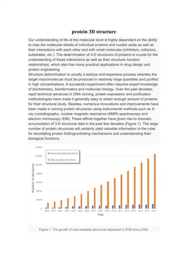
Practicals How many NEW structures were deposited in 2010 vs 2011? Hint: try this at PDBe vs RCSB PDB Much easier at RCSB PDB: http://www.rcsb.org/pdb/static.do?p=general_information/pdb_statistics/index.html http://www.rcsb.org/pdb/statistics/contentGrowthChart.do?content=total&seqid=100
Practicals How many structures determined by X-ray crystallography, NMR, Electron microscopy? Use Advanced Query at RCSB PDB
Find the structure with the highest resolution at RCSB PDB http://www.rcsb.org/pdb/statistics/histogram.do?mdcat=refine&mditem=ls_d_res_high&minLabel=0&maxLabel=5&numOfbars=10&name=Resolution
How many structures determined by X-ray crystallography, NMR, Electron microscopy? Use advanced Query at PDBe http://www.ebi.ac.uk/pdbe-srv/view/search?search_type=advanced
At RCSB PDB, find structures with imatinib as ligand. How many structures contain this ligand? What type of proteins contain this ligand? Use advanced query. Verify using the PDB ID for imatinib=STI Answer: Protein kinases
Amongst proteins with bound imatinib, there are structures for p38. http://www.rcsb.org/pdb/explore/explore.do?structureId=3HEC Find the official protein and gene name of p38 Most easily done via the UniProt entry Find the UniProt AC for p38.
Find structures for a human protein kinase with bound ADP and a resolution between 2 and 2.5 A: Use main query window, find structures with ligand ADP, then use options (Species, Resolution, Enzyme classification, …) to filter the result
Identifiy the residues that form hydrogen bonds with the product, via PDBsum and from the RCSB PDB website: e.g. 2IJM http://www.ebi.ac.uk/thornton-srv/databases/cgi-bin/pdbsum/RunLigplot.pdf?pdb=2ijm&pdf=YES&file=ligplot02_01 and Ligand Explorer from RCSB PDB (shown below)
At RCSB PDB, find structures for proteins similar to p38 Query: p38 imatinib in main search window, choose 3HEC: You get kinases
find structures for proteins similar to p38 via the DALI database (FSSP link on PDBsum page). What kind of proteins do you recover? http://ekhidna.biocenter.helsinki.fi/dali_server/results/3hec/index.html
Use the sequence of human PLK4 to find a matching structure (advanced search tools) Sequence: http://www.uniprot.org/uniprot/O00444.fasta
Look at protein structure classification in CATH and SCOP (e.g. via links for 3BRB from PDB or PDBsum). The aim is essentially to see that there are quantitative means to classify domain structures No hit in SCOP, so try keyword search, or start at top of hierarchy http://scop.mrc-lmb.cam.ac.uk/scop/search.cgi?ver=1.75&key=kinase&search_type=scop
How many PDB structures are represented in CATH and in SCOP? What is the reason for the difference relative to the total number of experimental structures? Check their home pages: http://www.cathdb.info/ http://scop.mrc-lmb.cam.ac.uk/scop/index.html More automated procedures can handle more data – human beings can handle less data, but using your brains adds extra value
Starting with the structure for human FTO, find other proteins with similar 3D-structure via PDB http://www.rcsb.org/pdb/explore/structureCluster.do?structureId=3LFM or DALI (FSSP) : http://ekhidna.biocenter.helsinki.fi/dali_server/results/3lfm/index.html What kind of proteins do you find (function, taxonomy)? Answer: dioxygenases, more specifically ALPHA-KETOGLUTARATE-DEPENDENT DIOXYGENASE Taxons: Mammalia (alkB homologs), and bacteria (alkB from E.coli) What are their ligands? Answer: alpha-ketoglutarate and iron http://www.rcsb.org/pdb/explore.do?structureId=2IUW
Do an alignment based on the 3D-structure and check if the ligand-binding residues are conserved. Do this with entries from closely related/different taxons. • Get the ligand-binding sites using PDBsum, UniProt annotation, or Ligand explorer • http://www.uniprot.org/uniprot/Q9C0B1#section_features • http://www.uniprot.org/uniprot/P05050#section_features • B) Get an alignment from RCSB PDB or DALI • http://www.rcsb.org/pdb/workbench/showPrecalcAlignment.do?action=pw_fatcat&mol=3LFM.A&mol=2IUW.A Very low sequence identity, but secondary structure is almost the same. Binding sites for catalytic iron (HxD and H) are boxed
Can you get the same proteins, doing a normal BLAST search with FTO? Answer: No, they are too different Have a look at the domain structure (InterPro) of FTO and the proteins that you identified. Compare InterPro lines: they are different http://www.uniprot.org/uniprot/P05050#section_x-ref http://www.uniprot.org/uniprot/Q9C0B1#section_x-ref
Find 3D-structures for transthyretin at RCSB PDB, then look for proteins with similar 3D-structure http://www.rcsb.org/pdb/explore/explore.do?structureId=1THC Similar structure: 2H6U What is the function of transthyretin, respectively of other proteins with similar structure? Have a look at the corresponding UniProt entries! How are they classified at CATH / at SCOP? Use text search http://scop.mrc-lmb.cam.ac.uk/scop/data/scop.b.c.d.e.b.b.html http://www.cathdb.info/cathnode/2.60.40.180 NB: the proteins are too similar to be distinguished by these means
More things to do (facultative) • Find protein structures with aspirin as ligand • Find the chemical structure for aspirin • Get all structures with the ligand aspirin. How many are there? • Go to CHEBI, get Smiles for aspirin • http://www.ebi.ac.uk/chebi/advancedSearchFT.do • http://www.ebi.ac.uk/chebi/searchId.do;E5D46C3E2363F473FCE4C201CC2078A6?chebiId=CHEBI:15365 • Go to RCSB PDB, use advanced search (smiles) • You can also try and draw “aspirin” using CHEBI