Download
1 / 25
260 likes | 581 Vues
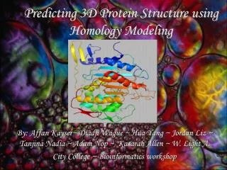
Predicting 3D Protein Structure using Homology Modeling. By: Affan Kayser~ Diadji Wague ~ Hua Yang ~ Jordan Liz ~ Tanjina Nadia ~ Adam Nop ~ Kasarah Allen ~ W. Light A. City College ~ Bioinformatics workshop. Introduction.

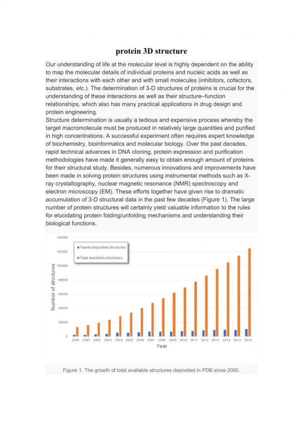
E N D
Predicting 3D Protein Structure using Homology Modeling By: Affan Kayser~ Diadji Wague ~ Hua Yang ~ Jordan Liz ~ Tanjina Nadia ~ Adam Nop ~ Kasarah Allen ~ W. Light A. City College ~ Bioinformatics workshop
Introduction Proteins play an important roles in various diseases. Through analyzing the properties and characteristics of proteins, we try to find their involvements in diseases. Structural analysis of the proteins pave our ways to find solutions to certain diseases. In order to analyze proteins which structures haven’t been determine yet, we use homology modeling to model proteins by using appropriate templates. MOE will help us do the homology modeling and be able to reveal the significances of the proteins in diseases from their structural analysis.
What is Homology Modeling? Homology modeling is based on the assumption that, if two proteins have similar sequences then they will have similar structures. If the sequence identity is greater than 90% then the predicted structure is practically the same as the template structure. If the sequence identity is lower than 25% then the predicted structure is not reliable. If the sequence identity is in the range 50%-80% then the homology modeling can generate reliable and informative structure. There are many software for homology modeling, the general procedure is: • Search for the template based on sequence similarity using Blast (an online software provided by the NCBI database) • Select the candidate template to make sure the E-value of the sequence alignment is less than 10-4 and sequence identity is greater than 50%. • Input the target sequence and the template sequence and structure into homology modeling software to do modeling, for example SWISS MODEL (http://swissmodel.expasy.org/SWISS-MODEL.html) ,or MOE. • Check the accuracy of the generated structure model.
MOE www.chemcomp.com • We are using MOE (molecular operating environment,) to do the modeling and this software will do all the steps for you when you input the sequences to be modeled. • We all found our protein sequences from the NCBI website and save in FASTA format. • We copied these sequences to MOE and searched for appropriate template. • After sequence alignment, we start to model the target protein structure based on the template structure. • Verify the local geometry of the generated model using Ramachandran Plot.
About TAL1…… • Tumor specific alteration of TAL1 occur in patients with T-Cell Acute Lymphoblastic Leukemia (T-ALL) • This alteration is caused by chromosomal rearrangement • Alteration of TAL1 may lead to leukemogenesis • Related to TAL2 and LYL1. Thus, has an etiologic role in leukemia
Continue……. • Contain a) amino acid residues 1-331 • b) truncated species ( residues 176-331) • Both have 1. basic helix-loop-helix motif; 2. protein dimerization; 3. DNA-binding domain that were found in transcription factors As products subgroup of bHLH protein which is a potential mediator of T-cell leukemogenesis
PDB ID: 1MDY: As template • TAL1 polypeptides do not have intrinsic DNA binding activity • can’t self-associate to form bHLH homodimers • May be able to interact with other bHLH protein in a stable manner • Two heterodimers(TAL1-E12 , TAL1-E47) that recognize E-box motif • structure model by MOE Loop Beta Helix E-box motif: a element found in various eukaryotic transcription enhancers
Ramachandran Plot It is a way to visualize dihedral angles φ against ψ of amino acid residues in protein structure
JunB • Psoriasis is an inflammatory disease of the skin and joints • JunB is an element of the AP-1 transcription factor and is known to coordinate cell differentiation, stress responses, cytokine expression in various organs and proliferation. But when a person develops psoriasis, the expression levels of JunB lessen.
This is the HIF1 protein structure done by MOE homology modeling. The PBD code: 1P97
Protein analysis of HIF1 • The predicted structure on the previous page displays the HIF1 protein, which is the main regulator of hypoxia-induced gene expression as well as the DNA binding protein. It is a heterodimeric transcription factor composed of HIF1A. • The HIF1 protein expressed by the structure concludes that it plays a big role in hypoxia. However, even though many proteins are known, this protein helps identify what hypoxia is really about since it does associate with the HIF1A gene.
Subtilisin Mutant • Acts as an Intramolecular Chaperone • Subtilisin-like serine Proteinase • It facilitates folding by acting as a template for its protease domain, which does not form A part of it. • There are two Subtilisin
Subtilisin Mutant cont. • Altered and wild type Subtilisin • They have identical amino-acid sequences • Differs in thermostability and substrate specificities. • Through a mutated intramoleculer chaperone: • An identical polypeptide can fold into an altered conformation • It also maintains memory of the folding process.
LDLR Structure • This structure was created by Moe. It’s the template for the LDLR protein. • This is the back bone to the structure of the LDL receptor protein.
Space Filling Model • Space filling molecular models show the relative atomic sizes of the atoms of the molecule. • This model shows the carbons (gray), hydrogen's (light gray), oxygen (blue), carbon monoxide's (red), and sulfur (yellow) of the protein.
LDL Receptor • Knowing the structure of the LDL receptor scientists can better understand the effects it has on Familial Hypercholesterolemia patients. • For example, scientist can find why the LDL receptor fails to activate and bind to the surface and can learn how to provide LDL receptors to FH patients.
What Is HER4? • HER4 , also known as ErbB4, is able to lower the expression level of breast and prostate tumors. • ErbB4 is a transmembrane receptor, which is a protein that covers the plasma membrane of the cell, that controls cell reproduction and differentiation. • It is apart of the HER family. • Has to be enzymatically separated which may change the function of its Intercellular Domain(ICD) as stated in several reports
Conclusion • The predicted structures of our target proteins help us analyze the significances of the proteins and its involvements in diseases. Thus, we have better understandings of the diseases.
Further Research We will continue to create models of unstructured proteins using modeling softwares other then MOE to see if they yield similar results.
References • http://www.epitomics.com/products/1200-1.htm • lhttp://www.ncbi.nlm.nih.gov/entrez/viewer.fcgi?db=protein&val=66393602 • http://www.ncbi.nlm.nih.gov/BLAST/Blast.cgiR • http://breast-cancer-research.com/content/8/2/R19 • http://www.findarticles.com/p/articles/mi_m0DFY/is_03_2001/ai_70738971 • MOE- Molecular Operating Environment • http://www.ncbi.nlm.nih.gov/entrez/viewer.fcgi?db=protein&val=31077211 • http://www.wrongdiagnosis.com/medical/hif1_protein.htm • http://www.genecards.org/cgi-bin/carddisp.pl?gene=HIF1A • Bacterial lipopolysaccharide induces HIF-1 activation in human monocytes via p44/42 MAPK and NF-kappaB.Frede S, Stockmann C, Freitag P, Fandrey J. • Wegele H, Muller L, Buchner J. (2004). Hsp70 and Hsp90 - a relay team for protein folding. Rev Physiol Biochem Pharmacol 151:1-44 • uhttp://www.ncbi.nlm.nih.gov/BLAST/Blast.cgi • uhttp://www.ebi.ac.uk/cgi-bin/clustalw/result?tool=clustalw&jobid=clustalw-20060809-16204016&treendisp=hide&treetype=new&sortby=seqno&color=yes • uhttp://www.ncbi.nlm.nih.gov/gquery/gquery.fcgi • uhttp://ad2004.com/Biblecodes/Hebrewmatrix/leukemia.html • http://www.humpath.com/IMG/jpg/psoriasis_nail_20xpasd-2.jpg • http://www.nature.com/nature/journal/v437/n7057/full/nature03963.html • http://www.nycornell.org/dermatology/research/img/psoriasis.jpg • http://www.chemcomp.com/software-pro.htm
Acknowledgements Special Thanks To: • Dr. Sat Bhattacharya • Dr. Yuying Q. Gosser • Joe Wu • Harlem Children Society • Bioinformatics workshop at City College of New York