Respiratory System: Functions and Anatomy
Learn about the respiratory system's role in providing oxygen to the body and eliminating carbon dioxide. Explore the functional anatomy of major respiratory organs like the nose, larynx, and lungs. Discover the processes of respiration, from pulmonary ventilation to gas exchange in the alveoli.

Respiratory System: Functions and Anatomy
E N D
Presentation Transcript
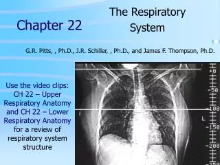
CHAPTER 22 1 THE RESPITORY SYSTEM PART A CHAPTER 22 Read pages: 804-819
Respiration 2 • Function: Supply the body with oxygen and dispose of carbon dioxide • Involves both the respiratory and the circulatory systems • Four processes that supply the body with O2 and dispose of CO2
Respiration 3 • Pulmonary ventilation (breathing):movement of air into and outof the lungs • External respiration: O2 and CO2exchange between the lungsand the blood • Transport: O2 and CO2in the blood • Internal respiration: O2 and CO2exchange between systemic bloodvessels and tissues Respiratory system Circulatory system
Respiratory System: Functional Anatomy 4 • Major organs • Nose, nasal cavity, and paranasal sinuses • Pharynx • Larynx • Trachea • Bronchi and their branches • Lungs and alveoli
Fig. 22.1 pg: 805 Nasal cavity Oral cavity Nostril Pharynx Larynx Left main (primary) bronchus Trachea Carina of trachea Right main (primary) bronchus Left lung Diaphragm Right lung Figure 21.1
Functional Anatomy 6 • Respiratory zone: site of gas exchange • Microscopic structures: respiratory bronchioles, alveolar ducts, and alveoli • Conducting zone: conduits to gas exchange sites • Includes all other respiratory structures • Respiratory muscles: diaphragm and other muscles that promote ventilation PLAY Animation: Rotatable face
The Nose 7 • Functions • Provides an airway for respiration • Moistens and warms the entering air • Filters and cleans inspired air • Serves as a resonating chamber for speech • Houses olfactory receptors
The Nose 8 • Two regions: external nose and nasal cavity • External nose: root, bridge, dorsum nasi, and apex • Nostrils (nares): bounded laterally by the alae
9. Fig. 22a pg: 806 Epicranius, frontal belly Root and bridge of nose Dorsum nasi Ala of nose Apex of nose Naris (nostril) Philtrum (a) Surface anatomy Figure 21.2a
Frontal bone 10. Fig 22b pg: 806 Nasal bone Septal cartilage Maxillary bone (frontal process) Lateral process of septal cartilage Minor alar cartilages Dense fibrous connective tissue Major alar cartilages (b) External skeletal framework Figure 21.2b
The Nose 11 • Nasal cavity: in and posterior to the external nose • Divided by a midline nasal septum • Posterior nasal apertures (choanae) open into the nasal pharynx koe.an’.e/funnel • Roof: ethmoid and sphenoid bones • Floor: hard and soft palates
Nasal Cavity 12 • Vestibule: nasal cavity superior to the nostrils • Hairs filter coarse particles from inspired air • Olfactory mucosa • Lines the superior nasal cavity • Contains smell receptors
Nasal Cavity 13 • Respiratory mucosa • Pseudostratified ciliated columnar epithelium • Mucous and serous secretions contain lysozyme (antibacterial enzyme) and defensins (natural antibiotic) • Cilia move contaminated mucus posteriorly to throat • Inspired air is warmed by plexuses of capillaries and veins • Sensory nerve endings triggers sneezing
Fig. 22.3c Pg: 808 14 Cribriform plate of ethmoid bone Frontal sinus Sphenoid sinus Nasal cavity Posterior nasal aperture Nasal conchae (superior, middle and inferior) Nasopharynx Pharyngeal tonsil Nasal meatuses (superior, middle, and inferior) Opening of pharyngotympanic tube Nasal vestibule Uvula Nostril Oropharynx Hard palate Palatine tonsil Soft palate Isthmus of the fauces Tongue Lingual tonsil Laryngopharynx Hyoid bone Larynx Epiglottis Vestibular fold Esophagus Thyroid cartilage Vocal fold Cricoid cartilage Trachea Thyroid gland (c) Illustration Figure 21.3c
Nasal Cavity 15 • Superior, middle, and inferior nasal conchae (Kong’.ke) • Protrude from the lateral walls • Increase mucosal area • Enhance air turbulence
Functions of the Nasal Mucosa and 16 Conchae • During inhalation, the conchae and nasal mucosa • Filter, heat, and moisten air • During exhalation these structures • Reclaim heat and moisture
Paranasal Sinuses 17 • In frontal, sphenoid, ethmoid, and maxillary bones • Lighten the skull and help to warm and moisten the air
Pharynx 18 • Muscular tube that connects to the • Nasal cavity and mouth superiorly • Larynx and esophagus inferiorly • From the base of the skull to the level of the sixth cervical vertebra
Fig. 22.3 pg. 808 Pharynx Nasopharynx Oropharynx Laryngopharynx (b) Regions of the pharynx Figure 21.3b
Nasopharynx 20 • Air passageway posterior to the nasal cavity • Lining: pseudostratified columnar epithelium • Soft palate and uvula close nasopharynx during swallowing • Pharyngeal tonsil (adenoids) on posterior wall • Pharyngotympanic (auditory) tubes open into the lateral walls
Oropharynx 21 • Passageway for food and air from the level of the soft palate to the epiglottis • Lining of stratified squamous epithelium • Palatine tonsils in the lateral walls of fauces • Lingual tonsil on the posterior surface of the tongue
Laryngopharynx 22 • Passageway for food and air • Posterior to the upright epiglottis • Extends to the larynx, where it is also continuous with the esophagus
Fig. 22.3c Pg. 808 Cribriform plate of ethmoid bone Frontal sinus Sphenoid sinus Nasal cavity Posterior nasal aperture Nasal conchae (superior, middle and inferior) Nasopharynx Pharyngeal tonsil Nasal meatuses (superior, middle, and inferior) Opening of pharyngotympanic tube Nasal vestibule Uvula Nostril Oropharynx Hard palate Palatine tonsil Soft palate Isthmus of the fauces Tongue Lingual tonsil Laryngopharynx Hyoid bone Larynx Epiglottis Vestibular fold Esophagus Thyroid cartilage Vocal fold Cricoid cartilage Trachea Thyroid gland (c) Illustration Figure 21.3c
Larynx 24 • Attaches to the hyoid bone and opens into the laryngopharynx • Continuous with the trachea • Functions • Provides a patent airway • Routes air and food into proper channels • Voice production
Larynx 25 • Cartilages of the larynx • Hyaline cartilage except for the epiglottis • Thyroid cartilage with laryngeal prominence (Adam’s apple) • Epiglottis: elastic cartilage; covers the laryngeal inlet during swallowing
Larynx 26 • Vocal ligaments • Contain elastic fibers • Form core of vocal folds (true vocal cords) • Opening between them is the glottis • Folds vibrate to produce sound as air rushes up from the lungs
Larynx 27 • Vestibular folds (false vocal cords) • Superior to the vocal folds • No part in sound production • Help to close the glottis during swallowing
Fig. 22.5 a&b pg. 811 Base of tongue Epiglottis Vestibular fold (false vocal cord) Vocal fold (true vocal cord) Glottis Inner lining of trachea Cuneiform cartilage Corniculate cartilage (a) Vocal folds in closed position; closed glottis (b) Vocal folds in open position; open glottis Figure 21.5
Voice Production 29 • Speech: intermittent release of expired air while opening and closing the glottis • Pitch is determined by the length and tension of the vocal cords • Loudness depends upon the force of air • Chambers of pharynx, oral, nasal, and sinus cavities amplify and enhance sound quality • Sound is “shaped” into language by muscles of the pharynx, tongue, soft palate, and lips
Larynx 30 • Vocal folds may act as a sphincter to prevent air passage • Example: Valsalva’s maneuver • Glottis closes to prevent exhalation • Abdominal muscles contract • Intra-abdominal pressure rises • Helps to empty the rectum or stabilizes the trunk during heavy lifting
Trachea 31 • Windpipe: from the larynx into the mediastinum • Wall composed of three layers • Mucosa: ciliated pseudostratified epithelium with goblet cells • Submucosa: connective tissue with seromucous glands • Adventitia: outermost layer made of connective tissue that encases the C-shaped rings of hyaline cartilage
Trachea 32 • Trachealis muscle • Connects posterior parts of cartilage rings • Contracts during coughing to expel mucus • Carina • Last tracheal cartilage • Point where trachea branches into two bronchi
Fig: 22.6a Pg. 812 Posterior Mucosa Esophagus Submucosa Trachealis muscle Seromucous gland in submucosa Lumen of trachea Hyaline cartilage Adventitia Anterior (a) Cross section of the trachea and esophagus Figure 21.6a
Fig: 22.6b Pg. 812 Mucosa • Pseudostratified ciliated columnar epithelium • Lamina propria (connective tissue) Submucosa Seromucous gland in submucosa Hyaline cartilage (b) Photomicrograph of the tracheal wall (320x) Figure 21.6b
Bronchi and Subdivisions 35 • Air passages undergo 23 orders of branching • Branching pattern called the bronchial (respiratory) tree
Conducting Zone Structures 36 • Trachea right and left main (primary) bronchi • Each main bronchus enters the hilum of one lung • Each main bronchus branches into lobar (secondary) bronchi (three right, two left) • Each lobar bronchus supplies one lobe
Conducting Zone Structures 37 • Each lobar bronchus branches into segmental (tertiary) bronchi • Segmental bronchi divide repeatedly • Bronchioles are less than 1 mm in diameter • Terminal bronchioles are the smallest, less than 0.5 mm diameter
Fig: 22.7 pg. 813 Trachea Superior lobe of left lung Left main (primary) bronchus Superior lobe of right lung Lobar (secondary) bronchus Segmental (tertiary) bronchus Middle lobe of right lung Inferior lobe of left lung Inferior lobe of right lung Figure 21.7
Conducting Zone Structures 39 • From bronchi through bronchioles, structural changes occur • Cartilage rings give way to plates; cartilage is absent from bronchioles • Epithelium changes from pseudostratified columnar to cuboidal; cilia and goblet cells become sparse • Relative amount of smooth muscle increases
Respiratory Zone 40 • Respiratory bronchioles, alveolar ducts, alveolar sacs (clusters of alveoli) • ~300 million alveoli account for most of the lungs’ volume and are the main site for gas exchange
Fig: 22.8a pg. 814 Alveoli Alveolar duct Respiratory bronchioles Alveolar duct Terminal bronchiole Alveolar sac (a) Figure 21.8a
Fig: 22.8b pg. 814 Emphysema/Chronic Obstructive Pulmonary Disease Respiratory bronchiole Alveolar duct Alveolar pores Alveoli Alveolar sac (b) Figure 21.8b
Respiratory Membrane 43 • ~0.5-m-thick air-blood barrier • Alveolar and capillary walls and their fused basement membranes • Alveolar walls • Single layer of squamous epithelium (type I cells) • Scattered type II cuboidal cells secrete surfactant and antimicrobial proteins
44 Fig. 22.9a pg. 816 Terminal bronchiole Respiratory bronchiole Smooth muscle Elastic fibers Alveolus Capillaries (a) Diagrammatic view of capillary-alveoli relationships Figure 21.9a
45 Fig. 22.9b pg. 816 Figure 21.9b
Alveoli 46 • Surrounded by fine elastic fibers • Contain open pores that • Connect adjacent alveoli • Allow air pressure throughout the lung to be equalized • House alveolar macrophages that keep alveolar surfaces sterile
47 Fig. 22.9c pg. 816 Red blood cell Nucleus of type I (squamous epithelial) cell Alveolar pores Capillary O2 Capillary Type I cell of alveolar wall CO2 Alveolus Macrophage Alveolus Endothelial cell nucleus Alveolar epithelium Fused basement membranes of the alveolar epithelium and the capillary endothelium Respiratory membrane Red blood cell in capillary Alveoli (gas-filled air spaces) Type II (surfactant- secreting) cell Capillary endothelium (c) Detailed anatomy of the respiratory membrane Figure 21.9c
Lungs 48 • Occupy all of the thoracic cavity except the mediastinum • Root: site of vascular and bronchial attachments • Costal surface: anterior, lateral, and posterior surfaces
49 Fig. 22.10c pg 817 Esophagus (in mediastinum) Posterior Vertebra Root of lung at hilum Right lung Parietal pleura • Left main bronchus • Left pulmonary artery • Left pulmonary vein Visceral pleura Left lung Pleural cavity Thoracic wall Pulmonary trunk Pericardial membranes Heart (in mediastinum) Anterior mediastinum Sternum Anterior (c) Transverse section through the thorax, viewed from above. Lungs, pleural membranes, and major organs in the mediastinum are shown. Figure 21.10c
Lungs 50 • Apex: superior tip • Base: inferior surface that rests on the diaphragm • Hilum: on mediastinal surface; site for attachment of blood vessels, bronchi, lymphatic vessels, and nerves • Cardiac notch of left lung: concavity that accommodates the heart