Download
1 / 1
10 likes | 123 Vues
Developing Novel Supported Membrane Interfaces for SPR Study of Transmembrane Proteins Heather Ferguson*, Matthew J. Linman † , Quan Cheng † † Department of Chemistry - University of California Riverside 92521 *Walla Walla University, WA 99324. OPTIONAL LOGO HERE. Results. Introduction.

E N D
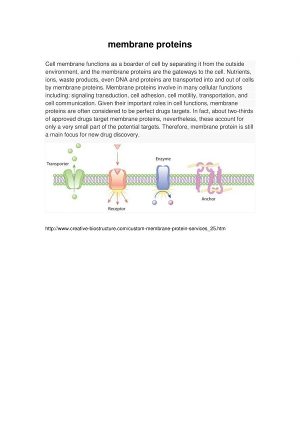
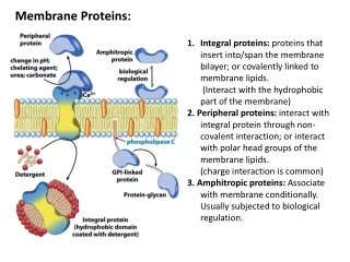
Developing Novel Supported Membrane Interfaces for SPR Study of Transmembrane Proteins Heather Ferguson*, Matthew J. Linman†, Quan Cheng† †Department of Chemistry - University of California Riverside 92521 *Walla Walla University, WA 99324 OPTIONALLOGO HERE Results Introduction Conclusion Membrane proteins are known key molecules in cellular activities such as maintaining cell structure, transport, and signalling. Therefore an understanding of the function, especially of the affinity property of membrane proteins, is of particular importance to the pharmaceutical industry where 60% of drug targets are membrane proteins.1 However, membrane proteins are difficult to study in their native environment and do not function properly when removed from a lipid membrane. We set out to mimic a mammalian lipid membrane environment using phosphatidylcholine (PC) vesicles that surround the transmembrane protein epidermal growth factor receptor (EGFR). EGFR is often overexpressed in cells in certain types of cancers such as breast, colon, and lung.2 Surface plasmon resonance (SPR) is employed to characterize interactions between the membrane protein and immobilized anti-EGFR TK, an antibody against the EGFR tyrosine kinase (TK) domain. Several surface chemistries have been examined in an effort to find a reproducible and functional biomimetic surface containing EGFR. An important step to the preparation of the sensing layers is successfully biotinylating proteins. These data show that BSA biotinylated with Amine-PEG3 (Figure 4) gave a much greater SPR signal than the non-biotinylated control response (Figure 3). • Biotinylation procedure needs to be perfected until it is more reliable and gives reproducible results. • Overexpressed EGFR cells show preferential binding to mAb for EGFR, Erbitux, compared to control cells. • Detected a slightly higher signal between cells over-expressing EGFR when using polyclonal anti-EGFR TK compared to control cells. Using a higher concentration of antibody may increase the analytical signal. • Current method of combining EGFR and PC vesicles can be improved. Figure 3.Sensorgram showing the response between unbiotinylated BSA and NeutrAvidin Figure 4. Sensorgram showing the response between Amine-PEG3-Biotinylated BSA and NeutrAvidin Future Work • Determine ideal membrane interface design for effective and functional EGFR immobilization for protein binding. • Create an interface based on the calcinated chip (glassified layer on gold) for direct immobilization of the EGFR in a membrane. 4 • Apply best interface design to a microarray format for high-throughput screening with SPR imaging. Methods Surface plasmon resonance is a label-free biosensing technique that is capable of real-time monitoring of biological interactions. P-polarized light excites the electrons in the gold layer, and results in a minimum of reflected light intensity, marking the surface plasmon angle. SPR is sensitive to changes in the index of refraction near the surface (< 200 nm). As molecules bind to the surface, the signal increases proportionally to the amount of accumulated mass. Figure 5.Sensorgram showing enhanced signal to mAb for EGFR, Erbitux, in the presence of overexpressed EGFR cells compared to control cells. References • Hopkins, A. L.; Groom, C. R. Nat.Rev. Drug Discovery 2002, 1, 727–730. • Hubbard, S. R. Cancer Cell 2005, 7, 287-288. • Li, Shiqing; et al. Cancer Cell 2005, 7, 301-311. • Linman, M. J.; Culver S.P.; Cheng Q. Langmuir 2009, 25, 3075-3082. Figure 1. Schematic of SPR set-up. The analyte in the flow channel are cells expressing EGFR and the immobilized molecule is anti-EGFR. Diagram from : Cooper, M.A. Nat. Rev. Drug Discovery 2002, 1, 517. Figure 2.Cartoon representation of the desired molecular layers on gold. Acknowledgements National Science Foundation BRITE REU program Cheng Lab Group Lipids cannot bind directly to gold, so a protein layer such as bovine serum albumin is used. NeutrAvidin and biotin bind together very strongly and serve as linking units to tether antibody and the lipid bilayer containing EGFR to the surface. Figure 6. Sensorgram showing the binding responses of control cells and cells overexpressing EGFR to anti-EGFR TK immobilized on a functionalized self-assembled monolayer (SAM). The signal for the EGFR-expressing cells is slightly higher than the control cells. Figure 7. Sensorgram showing the binding response between Erbitux and EGFR expressing cells combined with PC lipid membrane. Erbitux is a monoclonal antibody specific to an extracellular domain of EGFR.3 The lack of a strong signal indicates that the EGFR is not properly oriented in the membrane.