OSTEOLOGY BONES
OSTEOLOGY BONES. 23. September 2011 Wednesday. Kaan Yücel M.D., Ph.D . Osteology ( Gk , osteon, bone, logos, science) is the branch of medicine concerned with the development and diseases of bone tissue. The human skeleton 206 bones in adults .

OSTEOLOGY BONES
E N D
Presentation Transcript
OSTEOLOGY BONES 23. September2011Wednesday Kaan Yücel M.D., Ph.D.
Osteology (Gk, osteon, bone, logos, science) is the branch of medicine concerned with the development and diseases of bone tissue. The human skeleton 206 bones in adults
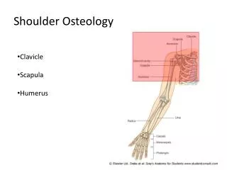
The skeletal system may be divided into • 2functional parts: • The axial skeleton • head (cranium or skull) • neck (hyoid bone and cervical vertebrae) • trunk (ribs, sternum, vertebrae, and sacrum) • The appendicular skeleton • Limbs • including those forming the shoulde & pelvic girdles
Bone is one of the hardest structures of the animal body, because of the calcification of its extracellular matrix. • Living bones have some elasticity (results from the organic matter) and great rigidity (results from their lamellous structures and tubes of inorganic calcium phosphate). • Its color, in a fresh state, is pinkish-white externally, and deep red within.
Cartilage and Bones • The skeleton is composed of cartilages and bones. • Cartilage is a resilient, semirigid form of connective tissue that forms parts of the skeleton where more flexibility is required—for example, where the costal cartilages attach the ribs to the sternum. • Also, the articulating surfaces (bearing surfaces) of bones participating in a synovial joint are capped with articular cartilage that provides smooth, low-friction, gliding surfaces for free movement.
Blood vessels do not enter cartilage (i.e., it is avascular); consequently, its cells obtain oxygen and nutrients by diffusion. • The proportion of bone and cartilage in the skeleton changes as the body grows; the younger a person is, the more cartilage he or she has. • The bones of a newborn are soft and flexible because they are mostly composed of cartilage.
Bone has a protective function; the skull and vertebral column, for example, protect the brain and spinal cord from injury; the sternum and ribs protect the thoracic and upper abdominal viscera. • It serves as a lever, as seen in the long bones of the limbs, and as an important storage area for calcium salts. • It houses and protects within its cavities the delicate blood-forming bone marrow.
Classification of Bones • Bones are classified according to their shape. • Long bones • Short bones • Flat bones • Irregular bones • Sesamoid bones
Classification of Bones • Long bones are tubular (e.g., the humerus in the arm).
Classification of Bones • Short bones are cuboidal and are found only in the tarsus (ankle) and carpus (wrist).
Classification of Bones • Flat bones usually serve protective functions (e.g., the flat bones of the cranium protect the brain).
Classification of Bones • Irregular bones have various shapes other than long, short, or flat (e.g., bones of the face).
Classification of Bones • Sesamoidbones(e.g., the patella or knee cap) develop in certain tendons and are found where tendons cross the ends of long bones in the limbs; they protect the tendons from excessive wear and often change the angle of the tendons as they pass to their attachments.
There are two types of bones according to histological features: compact bone and spongy (trabecular) bone. • They are distinguished by the relative amount of solid matter and by the number and size of the spaces they contain.
All bones have a superficial thin layer of compact bone around a central mass of spongy bone, except where the latter is replaced by a medullary (marrow) cavity. • Spongy bone is found at theexpanded heads of long bones and fills most irregular bones. • Compact bone forms the outer shell of all bonesand also the shafts in long bones.
Bone Markings and Formations • Bone markings appear wherever tendons, ligaments, and fascias are attached or where arteries lie adjacent to or enter bones. • Other formations occur in relation to the passage of a tendon (often to direct the tendon or improve its leverage) or to control the type of movement occurring at a joint.
Bone Markings and Formations • Surfaces of the bones are not smooth. • Bones display elevations, depressions and holes. • The surface features on the bones are given names to distinguish and define them.
Vasculature and Innervation of Bones • Bones are richly supplied with blood vessels. • Veins accompany arteries. • Nerves accompany blood vessels supplying bones.
The skull is supported on the summit of the vertebral column, and is of an oval shape, wider behind than in front. It is composed of a series of flattened or irregular bones which, with one exception (the mandible), are immovably jointed together. It is divisible into two parts: cranium, which lodges and protects the brain, consists of 8 bones skeleton of the face,of 14
Ossa Cranii The Occipital bone: situated at the back and lower part of the cranium, is trapezoid in shape and curved on itself. It is pierced by a large oval aperture, the foramen magnum, through which the cranial cavity communicates with the vertebral canal.
The curved, expanded plate behind the foramen magnum is named the squama; the thick, somewhat quadrilateral piece in front of the foramen is called the basilar part, whilst on either side of the foramen is the lateral portion.
Some prominent features of the occipital bone: External occipital protuberance: between the summit of the bone and the foramen magnum Nuchal lines: Lateral to the external occipital protuberance Cruciate eminence: Divides the interior surface of the occipital bone into four fossae.
Some prominent features of the occipital bone: Internal occipital protuberance:At the point of intersection of the four divisions of the cruciate eminence Internal occipital crest:The lower division of the cruciate eminence
The Parietal Bones: form, by their union, the sides and roof of the cranium. • Each bone is irregularly quadrilateral in form. • The external surface is convex, smooth, and marked near the center by an eminence, the parietal eminence (tuber parietale). • Crossing the middle of the bone in an arched direction are two curved lines, the superior and inferior temporal lines.
The Frontal Bone: resembles a cockle-shell in form, and consists of two portions: Avertical portion, the squama, corresponding with the region of the forehead An orbital or horizontal portion, which enters into the formation of the roofs of the orbital and nasal cavities.
Some prominent features of the frontal bone: Nasal process:The downward projection of the nasal part of the frontal bone which terminates as the nasal spine. Frontal crest:The internal surface of the squamafrontalis of the frontal bone is concave and presents in the upper part of the middle line a vertical groove, the sagittal sulcus, the edges of which unite below to form a ridge, the frontal crest. Zygomatic process:is the part of the zygomatic process consisting of the frontal bone.
Some prominent features of the frontal bone: Foramen cecum:The frontal crest of the frontal bone ends below in a small notch which is converted into a foramen, the foramen cecum (or foramen caecum), by articulation with the ethmoid.
The Temporal Bones: are situated at the sides and base of the skull. Each consists of five parts, viz., the squama, the petrous, mastoid, and tympanic parts, and the styloid process. Some prominent features of the temporal bone: Zygomatic process: projects from the lower part of the squama as a long, arched process.
The Sphenoid Bone: is situated at the base of the skull in front of the temporals and basilar part of the occipital. • It somewhat resembles a bat with its wings extended, and is divided into a median portion or body, two great and two small wings extending outward from the sides of the body, and two pterygoid processes which project from it below.
Tuberculumsellæ:behind the chiasmatic groove is an elevation, the tuberculumsellae; and still more posteriorly, a deep depression, the sellaturcica, the deepest part of which lodges the hypophysiscerebri and is known as the fossa hypophyseos(or fossa hypophysialis).
Clivus:(Latin for "slope") is a part of the cranium, a shallow depression behind the dorsum sellæ that slopes obliquely backward.
The Ethmoid bone: is exceedingly light and spongy, and cubical in shape. • It is situated at the anterior part of the base of the cranium, between the two orbits, at the roof of the nose, and contributes to each of these cavities.
Theethmoid bone consists of 4parts: • Ahorizontal or cribriform plate, forming part of the base of the cranium; • Aperpendicular plate, constituting part of the nasal septum; • 2lateral masses or labyrinths
Cranial Fossas • The inferior and anterior parts of the frontal lobes of the brain occupy the anterior cranial fossa, the shallowest of the three cranial fossae. • The fossa is formed by the frontal bone anteriorly, the ethmoid bone in the middle, and the body and lesser wings of the sphenoid posteriorly.
The butterfly-shaped middle cranial fossa has a central part composed of the sellaturcica on the body of the sphenoid and large, depressed lateral parts on each side.
The posterior cranial fossa, the largest and deepest of the three cranial fossae, lodges the cerebellum, pons, and medulla oblongata. • The posterior cranial fossa is formed mostly by the occipital bone.
The Facial Bones Nasal Bones Maxillæ(Upper Jaw) 3. Lacrimal Bone 4. ZygomaticBone (Malar Bone) 5. Palatine Bone 6. Inferior Nasal Concha (Concha Nasalis Inferior; Inferior Turbinated Bone) 7. Vomer 8. Mandible (Lower Jaw) 9. Hyoid Bone
Vertebral column, ribs and the sternum
Ribs (L. costae) are curved, flat bones that form most of the thoracic cage. There are 3 types of ribs: • True (vertebrocostal) ribs (1st-7th ribs): • directly to the sternum. • False (vertebrochondral) ribs • (8th, 9th, and usually 10th ribs): • indirect withthesternum • Floating (vertebral, free) ribs • (11th, 12th, and sometimes 10th ribs): • No connectionwiththesternum
Typical ribs (3rd-9th) have the following components: • Head • Neck • Tubercle • Body (shaft) • .
Costal cartilages prolong the ribs anteriorly and contribute to the elasticity of the thoracic wall, providing a flexible attachment for their anterior ends.